Our official English website, www.x-mol.net, welcomes your feedback! (Note: you will need to create a separate account there.)
Comparative analysis of striatal [18F]FDOPA uptake in a partial lesion model of Parkinson's disease in rats: Ratio method versus graphical model
SYNAPSE ( IF 2.3 ) Pub Date : 2022-03-06 , DOI: 10.1002/syn.22231 Arturo Avendaño-Estrada 1 , Leticia Verdugo-Díaz 2 , Miguel A Ávila-Rodríguez 1
SYNAPSE ( IF 2.3 ) Pub Date : 2022-03-06 , DOI: 10.1002/syn.22231 Arturo Avendaño-Estrada 1 , Leticia Verdugo-Díaz 2 , Miguel A Ávila-Rodríguez 1
Affiliation
|
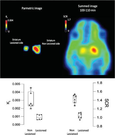
Animal models of Parkinson's disease are useful to evaluate new treatments and to elucidate the etiology of the disease. Hence, it is necessary to have methods that allow quantification of their effectiveness. [18F]FDOPA-PET (FDOPA-PET) imaging is outstanding for this purpose because of its capacity to measure changes in the dopaminergic pathway noninvasively and in vivo. Nevertheless, PET acquisition and quantification is time-consuming making it necessary to find faster ways to quantify FDOPA-PET data. This study evaluated Male Wistar rats by FDOPA, before and after being partially injured with 6-OHDA unilaterally. MicroPET scans with a duration of 120 min were acquired and Patlak reference plots were created to estimate the influx constant Kc in the striatum using the full dynamic scan data. Additionally, simple striatal-to-cerebral ratios (SCR) of short static acquisitions were computed and compared with the Kc values. Good correlation (r > 0.70) was obtained between Kc and SCR, acquired between 80–120 min after FDOPA administration with frames of 10 or 20 min and both methods were able to separate the FDOPA-uptake of healthy controls from that of the PD model (SCR −28%, Kc −71%). The present study concludes that Kc and SCR can be trustfully used to discriminate partially lesioned rats from healthy controls.
中文翻译:

大鼠帕金森病部分病变模型中纹状体 [18F]FDOPA 摄取的比较分析:比率法与图形模型
帕金森病的动物模型可用于评估新的治疗方法和阐明疾病的病因。因此,有必要采用能够量化其有效性的方法。[ 18 F]FDOPA-PET (FDOPA-PET) 成像非常适合此目的,因为它能够无创地和在体内测量多巴胺能通路的变化。然而,PET 采集和量化非常耗时,因此有必要找到更快的方法来量化 FDOPA-PET 数据。本研究通过 FDOPA 评估雄性 Wistar 大鼠在 6-OHDA 单方面部分损伤之前和之后。获得持续时间为 120 分钟的 MicroPET 扫描,并创建 Patlak 参考图以估计流入常数K c在纹状体中使用全动态扫描数据。此外,计算了短静态采集的简单纹状体与大脑比率 (SCR) 并与K c值进行比较。K c和 SCR 之间获得了良好的相关性 ( r > 0.70) ,在 FDOPA 给药后 80-120 分钟之间获得,帧为 10 或 20 分钟,两种方法都能够将健康对照的 FDOPA 摄取与 PD 的摄取分开模型(SCR -28%,K c -71%)。本研究得出结论,K c和 SCR 可以可靠地用于区分部分损伤的大鼠和健康对照。
更新日期:2022-03-06
中文翻译:
大鼠帕金森病部分病变模型中纹状体 [18F]FDOPA 摄取的比较分析:比率法与图形模型
帕金森病的动物模型可用于评估新的治疗方法和阐明疾病的病因。因此,有必要采用能够量化其有效性的方法。[ 18 F]FDOPA-PET (FDOPA-PET) 成像非常适合此目的,因为它能够无创地和在体内测量多巴胺能通路的变化。然而,PET 采集和量化非常耗时,因此有必要找到更快的方法来量化 FDOPA-PET 数据。本研究通过 FDOPA 评估雄性 Wistar 大鼠在 6-OHDA 单方面部分损伤之前和之后。获得持续时间为 120 分钟的 MicroPET 扫描,并创建 Patlak 参考图以估计流入常数K c在纹状体中使用全动态扫描数据。此外,计算了短静态采集的简单纹状体与大脑比率 (SCR) 并与K c值进行比较。K c和 SCR 之间获得了良好的相关性 ( r > 0.70) ,在 FDOPA 给药后 80-120 分钟之间获得,帧为 10 或 20 分钟,两种方法都能够将健康对照的 FDOPA 摄取与 PD 的摄取分开模型(SCR -28%,K c -71%)。本研究得出结论,K c和 SCR 可以可靠地用于区分部分损伤的大鼠和健康对照。


 京公网安备 11010802027423号