International Journal of Intelligent Robotics and Applications Pub Date : 2023-01-18 , DOI: 10.1007/s41315-022-00269-5 Avleen Malhi , Reaya Grewal , Husanbir Singh Pannu
|
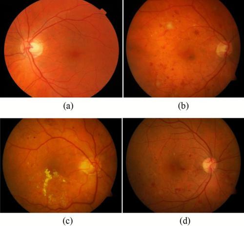
Diabetic Retinopathy is an eye disorder that affects people suffering from diabetes. Higher sugar levels in blood leads to damage of blood vessels in eyes and may even cause blindness. Diabetic retinopathy is identified by red spots known as microanuerysms and bright yellow lesions called exudates. It has been observed that early detection of exudates and microaneurysms may save the patient’s vision and this paper proposes a simple and effective technique for diabetic retinopathy. Both publicly available and real time datasets of colored images captured by fundus camera have been used for the empirical analysis. In the proposed work, grading has been done to know the severity of diabetic retinopathy i.e. whether it is mild, moderate or severe using exudates and micro aneurysms in the fundus images. An automated approach that uses image processing, features extraction and machine learning models to predict accurately the presence of the exudates and micro aneurysms which can be used for grading has been proposed. The research is carried out in two segments; one for exudates and another for micro aneurysms. The grading via exudates is done based upon their distance from macula whereas grading via micro aneurysms is done by calculating their count. For grading using exudates, support vector machine and K-Nearest neighbor show the highest accuracy of 92.1% and for grading using micro aneurysms, decision tree shows the highest accuracy of 99.9% in prediction of severity levels of the disease.
中文翻译:
使用数字视网膜图像检测和糖尿病视网膜病变分级
糖尿病性视网膜病变是一种影响糖尿病患者的眼部疾病。血液中较高的糖含量会导致眼睛血管受损,甚至可能导致失明。糖尿病性视网膜病变通过称为微动脉瘤的红色斑点和称为渗出物的亮黄色病变来识别。据观察,渗出液和微动脉瘤的早期检测可能会挽救患者的视力,本文提出了一种简单有效的糖尿病视网膜病变技术。眼底相机捕获的彩色图像的公开可用数据集和实时数据集均已用于实证分析。在拟议的工作中,已经完成分级以了解糖尿病性视网膜病变的严重程度,即使用眼底图像中的渗出液和微动脉瘤是轻度、中度还是重度。一种使用图像处理的自动化方法,已经提出了特征提取和机器学习模型来准确预测可用于分级的渗出液和微动脉瘤的存在。研究分两个部分进行;一种用于渗出液,另一种用于微动脉瘤。通过渗出物的分级是根据它们与黄斑的距离进行的,而通过微动脉瘤的分级是通过计算它们的数量来进行的。对于使用渗出液的分级,支持向量机和 K-最近邻显示出 92.1% 的最高准确度,对于使用微动脉瘤的分级,决策树在预测疾病的严重程度方面显示出 99.9% 的最高准确度。研究分两个部分进行;一种用于渗出液,另一种用于微动脉瘤。通过渗出物的分级是根据它们与黄斑的距离进行的,而通过微动脉瘤的分级是通过计算它们的数量来进行的。对于使用渗出液的分级,支持向量机和 K-最近邻显示出 92.1% 的最高准确度,对于使用微动脉瘤的分级,决策树在预测疾病的严重程度方面显示出 99.9% 的最高准确度。研究分两个部分进行;一种用于渗出液,另一种用于微动脉瘤。通过渗出物的分级是根据它们与黄斑的距离进行的,而通过微动脉瘤的分级是通过计算它们的数量来进行的。对于使用渗出液的分级,支持向量机和 K-最近邻显示出 92.1% 的最高准确度,对于使用微动脉瘤的分级,决策树在预测疾病的严重程度方面显示出 99.9% 的最高准确度。


 京公网安备 11010802027423号