Radiation and Environmental Biophysics ( IF 1.7 ) Pub Date : 2023-03-18 , DOI: 10.1007/s00411-023-01020-9 Yeşim Deniz 1 , Ezgi Işıktaş Acar 2 , Çiğdem Çetin Genç 3
|
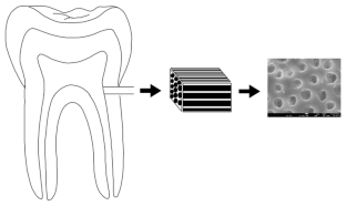
The aims of the study were to analyze the effects of therapeutic radiation on human root dentin samples from the aspect of possible alterations in crystallinity, micro-morphology, and composition. Fifty-six root dentin specimens were divided into seven groups (0, 10, 20, 30, 40, 50, and 60 Gy). Scanning electron microscope (SEM), energy-dispersive X-ray spectroscopy (EDX) and X-ray diffraction (XRD) analyses were performed on pulpal surfaces of root dentin after being irradiated by 6MV photon energy. Mineral compositions, Ca/P, P/N, Ca/N ratios, and hydroxyapatite pikes were calculated. Some deuteriations on the dentin surface were observed in SEM images after 30 Gy and subsequent doses. One-way ANOVA revealed that there was no significant alteration in weight percentages of C, O, Mg, Ca, P, and N between groups. Radiation did not influence stoichiometric Ca/P, Ca/N, and P/N molar ratios. XRD analysis did not show a remarkable decline in hydroxyapatite pikes by the increasing doses. Radiotherapy changes the micromorphology of circumpulpal dentin but does not affect elemental composition and crystallinity.
中文翻译:
直接治疗辐射对牙根牙本质牙髓表面的影响:一项体外研究
该研究的目的是从结晶度、微观形态和成分的可能改变方面分析治疗辐射对人根牙本质样本的影响。将 56 个牙根标本分为七组(0、10、20、30、40、50 和 60 Gy)。对经 6MV 光子能量照射后的根牙本质牙髓表面进行扫描电子显微镜 (SEM)、能量色散 X 射线光谱 (EDX) 和 X 射线衍射 (XRD) 分析。计算了矿物成分、Ca/P、P/N、Ca/N 比率和羟基磷灰石长矛。在 30 Gy 和后续剂量后,在 SEM 图像中观察到牙本质表面上的一些氘化。单向方差分析表明,各组之间 C、O、Mg、Ca、P 和 N 的重量百分比没有显着变化。辐射不影响化学计量的 Ca/P、Ca/N 和 P/N 摩尔比。XRD 分析未显示羟基磷灰石长矛因剂量增加而显着下降。放疗改变了牙髓周围牙本质的微观形态,但不影响元素组成和结晶度。


 京公网安备 11010802027423号