NanoImpact ( IF 4.9 ) Pub Date : 2023-04-28 , DOI: 10.1016/j.impact.2023.100465 Jasmin Kniese 1 , Sven Ritschar 2 , Lina Bünger 1 , Heike Feldhaar 3 , Christian Laforsch 3 , Andreas Römpp 1 , Heinar Schmidt 1
|
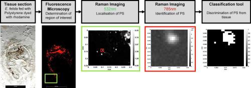
The uptake of microplastic particles (MPP) by organisms is frequently described and poses a potential risk for these organisms and ultimately for humans either through direct uptake or trophic transfer. Currently, the in-situ detection of MPP in organisms is typically based on histological examination of tissue sections after uptake of fluorescently-labelled MPP and is thus not feasible for environmental samples. The alternative approach is purification of MPP from whole organisms or organs by chemical digestion and subsequent spectroscopic detection (FT-IR or Raman). While this approach is feasible for un-labelled particles it goes along with loss of any spatial information related to the location in the tissue. In our study we aimed at providing a workflow for the localisation and identification of non-fluorescent and fluorescent polystyrene (PS) particles (fragments, size range 2–130 μm) in tissue sections of the model organism Eisenia fetida with Raman spectroscopic imaging (RSI). We provide methodological approaches for the preparation of the samples, technical parameters for the RSI measurements and data analysis for PS differentiation in tissue sections. The developed approaches were combined in a workflow for the in-situ analysis of MPP in tissue sections. The spectroscopic analysis requires differentiation of spectra of MPP and interfering compounds, which is challenging given the complexity of tissue. Therefore, a classification algorithm was developed to differentiate PS particles from haem, intestinal contents and surrounding tissue. It allows the differentiation of PS particles from protein in the tissue of E. fetida with an accuracy of 95%. The smallest PS particle detected in the tissue was 2 μm in diameter. We show that it is possible to localise and identify non-fluorescent and fluorescent ingested PS particles directly in tissue sections of E. fetida in the gut lumen and the adjacent tissue.
中文翻译:
使用拉曼光谱成像定位和识别组织切片中的聚苯乙烯颗粒
生物体对微塑料颗粒 (MPP) 的吸收经常被描述,并且通过直接吸收或营养转移对这些生物体并最终对人类构成潜在风险。目前,生物体中 MPP 的原位检测通常基于摄取荧光标记的 MPP 后组织切片的组织学检查,因此对于环境样品不可行。另一种方法是通过化学消化和随后的光谱检测(FT-IR 或拉曼)从整个生物体或器官中纯化 MPP。虽然这种方法对于未标记的粒子是可行的,但它会丢失与组织中位置相关的任何空间信息。具有拉曼光谱成像 (RSI) 的Eisenia fetida 。我们提供了样品制备方法、RSI 测量的技术参数和组织切片中 PS 分化的数据分析。开发的方法结合在一个工作流程中,用于组织切片中 MPP 的原位分析。光谱分析需要区分 MPP 和干扰化合物的光谱,鉴于组织的复杂性,这具有挑战性。因此,开发了一种分类算法来区分 PS 颗粒与血红素、肠内容物和周围组织。它可以区分 PS 颗粒和E. fetida组织中的蛋白质准确率为95%。在组织中检测到的最小 PS 颗粒直径为 2 μm。我们表明,可以直接在肠腔和邻近组织的E. fetida组织切片中定位和识别非荧光和荧光摄入的 PS 颗粒。


 京公网安备 11010802027423号