Forensic Chemistry ( IF 2.7 ) Pub Date : 2023-05-26 , DOI: 10.1016/j.forc.2023.100507 Kgalalelo Rampete , Colin I. Elliott , Theresa Stotesbury
|
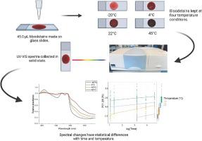
Determining the time since deposition (TSD) of bloodstained evidence can be an important process in forensic investigations. Hemoglobin is often examined as a biomolecule of interest for these purposes due to the known ex vivo oxidative changes to its structure. These time-dependent oxidative processes have previously been probed using UV–VIS spectroscopy following the resuspension of bloodstains. Our study investigated the solid-state VIS spectra of degrading bloodstains without sample pre-treatment, effectively bypassing the need for resuspension. A total of 128 bloodstains from eight biological replicates were created and stored on glass slides in four temperature conditions: −20 °C, 4 °C, 22 °C, and 45 °C (see graphical abstract, created with BioRender.com). Spectra were acquired from 380−800 nm at five time points spanning 96 h. The peak area of the methemoglobin (metHb) band displayed the largest time and temperature differences, an interesting contrast to previous literature using the Soret band for TSD. Principal Component Analysis (PCA) demonstrated that storage temperature delineated the data, with the metHb band showing the greatest contributions to PC1. Linear mixed models from the PCA data with time showed clear TSD relationships with temperature, and with minimal inter-donor variability. Overall, this work complements the UV–VIS analysis of bloodstains for TSD estimation, with the importance of noting clear differences between phases and sample preparation methods.
中文翻译:
监测降解血迹的固态 VIS 轮廓
确定血迹证据沉积后的时间 (TSD) 可能是法医调查中的一个重要过程。由于已知其结构的离体氧化变化,血红蛋白经常被作为用于这些目的的感兴趣的生物分子进行检查。先前已在血迹重新悬浮后使用紫外可见分光光度法探测了这些时间依赖性氧化过程。我们的研究研究了无需样品预处理即可降解血迹的固态 VIS 光谱,有效地绕过了重新悬浮的需要。总共 128 个来自 8 个生物复制品的血迹被创建并在四种温度条件下储存在载玻片上:−20 °C、4 °C、22 °C 和 45 °C(参见由 BioRender.com 创建的图形摘要))。在 96 小时内的五个时间点从 380−800 nm 处获取光谱。高铁血红蛋白 (metHb) 带的峰面积显示出最大的时间和温度差异,这与之前使用 Soret 谱带检测 TSD 的文献形成了有趣的对比。主成分分析 (PCA) 表明存储温度对数据进行了描述,其中 metHb 波段对 PC1 的贡献最大。PCA 数据随时间的线性混合模型显示了与温度的清晰 TSD 关系,并且捐赠者之间的变异性最小。总体而言,这项工作补充了用于 TSD 估计的血迹 UV-VIS 分析,并指出了阶段和样品制备方法之间明显差异的重要性。


 京公网安备 11010802027423号