Herz ( IF 1.7 ) Pub Date : 2023-07-25 , DOI: 10.1007/s00059-023-05198-y Meng Wang 1 , Xiaochen Wang 1 , Feng Gao 1 , Pei Bao 1 , Zheng Huang 1
|
Background
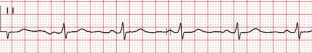
The P wave peak time (PWPT) is a predictor of paroxysmal atrial fibrillation (PAF). High-power short-duration ablation has been associated with improved durability of circumferential pulmonary vein electrical isolation (PVI). We investigated the effect of high-power short-duration PVI on PWPT in patients with PAF.
Methods
Out of 111 patients with PAF, 91 received radiofrequency ablation (ablation group) and 20 received medication treatment (control group). A VIZIGO sheath and an STSF catheter (Biosense Webster, CA, USA) were used together for high-power short-duration circumferential PVI at ablation index values of 500 and 400 for the anterior and posterior walls, respectively. The patients were followed up for 12 months.
Results
The preoperative PWPT in the ablation group was similar to that in the control group: PWPT II = 54.38 ± 6.18 ms vs. 54.35 ± 6.12 ms (p > 0.05), PWPT V1 = 54.19 ± 6.21 ms vs. 54.31 ± 6.08 ms (p > 0.05), respectively. Circumferential PVI was achieved for all patients in the ablation group during the operation. At the 12-month follow-up, there were seven cases of AF recurrence. The PWPT in the ablation group 12 months postoperatively was shorter than the preoperative value: PWPT II = 49.39 ± 7.11 ms vs. 54.38 ± 6.18 ms (p < 0.001), PWPT V1 = 47.69 ± 7.01 ms vs. 54.19 ± 6.21 ms (p < 0.001). The PWPT in the patients with AF recurrence was significantly longer than that in the non-recurrence patients: PWPT II = 50.48 ± 7.12 ms vs. 47.33 ± 6.21 ms (p < 0.001), PWPT V1 = 50.84 ± 7.05 ms vs. 47.19 ± 6.27 ms, (p < 0.001). The PWPT of the control group at the 12-month follow-up was similar to the baseline level: PWPT II = 54.32 ± 6.20 ms vs. 54.35 ± 6.12 ms (p > 0.05), PWPT V1 = 53.89 ± 6.01 ms vs. 54.31 ± 6.08 ms (p > 0.05).
Conclusion
The results showed that high-power short-duration PVI had a positive effect on PWPT, which is a predictor of PAF.
中文翻译:
高功率短时肺静脉隔离对 PWPT 的影响——阵发性心房颤动的预测因子
背景
P 波峰值时间 (PWPT) 是阵发性心房颤动 (PAF) 的预测因子。高功率短时消融与提高环肺静脉电隔离(PVI)的耐久性有关。我们研究了高功率短时 PVI 对 PAF 患者 PWPT 的影响。
方法
111 例 PAF 患者中,91 例接受射频消融(消融组),20 例接受药物治疗(对照组)。VIZIGO 护套和 STSF 导管(Biosense Webster,加利福尼亚州,美国)一起用于前壁和后壁消融指数值分别为 500 和 400 的高功率短持续时间圆周 PVI。对患者进行了为期 12 个月的随访。
结果
消融组术前 PWPT 与对照组相似:PWPT II = 54.38 ± 6.18 ms vs. 54.35 ± 6.12 ms ( p > 0.05),PWPT V 1 = 54.19 ± 6.21 ms vs. 54.31 ± 6.08 ms ( p > 0.05),分别。消融组所有患者术中均获得圆周PVI。12个月随访时,有7例房颤复发。消融组术后12个月的PWPT短于术前:PWPT II = 49.39 ± 7.11 ms vs. 54.38 ± 6.18 ms ( p < 0.001),PWPT V 1 = 47.69 ± 7.01 ms vs. 54.19 ± 6.21 ms ( p < 0.001)。房颤复发患者的 PWPT 明显长于未复发患者:PWPT II = 50.48 ± 7.12 ms vs. 47.33 ± 6.21 ms (p < 0.001),PWPT V 1 = 50.84 ± 7.05 ms vs. 47.19 ± 6.27 毫秒,(p < 0.001)。对照组12个月随访时的PWPT与基线水平相似:PWPT II = 54.32 ± 6.20 ms vs. 54.35 ± 6.12 ms ( p > 0.05),PWPT V 1 = 53.89 ± 6.01 ms vs. 54.31 ± 6.08 毫秒(p > 0.05)。
结论
结果表明,高功率短时 PVI 对 PWPT 有积极影响,而 PWPT 是 PAF 的预测因子。


 京公网安备 11010802027423号