Cardiovascular Engineering and Technology ( IF 1.8 ) Pub Date : 2023-08-31 , DOI: 10.1007/s13239-023-00679-x Scott MacDonald Black 1 , Craig Maclean 2 , Pauline Hall Barrientos 3 , Konstantinos Ritos 4, 5 , Asimina Kazakidi 1
|
Purpose
Segmentation and reconstruction of arterial blood vessels is a fundamental step in the translation of computational fluid dynamics (CFD) to the clinical practice. Four-dimensional flow magnetic resonance imaging (4D Flow-MRI) can provide detailed information of blood flow but processing this information to elucidate the underlying anatomical structures is challenging. In this study, we present a novel approach to create high-contrast anatomical images from retrospective 4D Flow-MRI data.
Methods
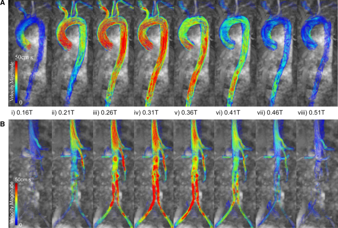
For healthy and clinical cases, the 3D instantaneous velocities at multiple cardiac time steps were superimposed directly onto the 4D Flow-MRI magnitude images and combined into a single composite frame. This new Composite Phase-Contrast Magnetic Resonance Angiogram (CPC-MRA) resulted in enhanced and uniform contrast within the lumen. These images were subsequently segmented and reconstructed to generate 3D arterial models for CFD. Using the time-dependent, 3D incompressible Reynolds-averaged Navier–Stokes equations, the transient aortic haemodynamics was computed within a rigid wall model of patient geometries.
Results
Validation of these models against the gold standard CT-based approach showed no statistically significant inter-modality difference regarding vessel radius or curvature (p > 0.05), and a similar Dice Similarity Coefficient and Hausdorff Distance. CFD-derived near-wall hemodynamics indicated a significant inter-modality difference (p > 0.05), though these absolute errors were small. When compared to the in vivo data, CFD-derived velocities were qualitatively similar.
Conclusion
This proof-of-concept study demonstrated that functional 4D Flow-MRI information can be utilized to retrospectively generate anatomical information for CFD models in the absence of standard imaging datasets and intravenous contrast.
中文翻译:
使用 4D 流 MRI 幅度图像的多个时间框架重建和验证计算流体动力学的动脉几何形状
目的
动脉血管的分割和重建是将计算流体动力学(CFD)转化为临床实践的基本步骤。四维流磁共振成像(4D Flow-MRI)可以提供血流的详细信息,但处理这些信息以阐明潜在的解剖结构具有挑战性。在这项研究中,我们提出了一种从回顾性 4D Flow-MRI 数据创建高对比度解剖图像的新方法。
方法
对于健康和临床病例,多个心脏时间步长的 3D 瞬时速度直接叠加到 4D Flow-MRI 幅度图像上,并组合成单个复合帧。这种新型复合相差磁共振血管造影 (CPC-MRA) 增强了管腔内的均匀对比度。随后对这些图像进行分割和重建,以生成 CFD 的 3D 动脉模型。使用随时间变化的 3D 不可压缩雷诺平均纳维-斯托克斯方程,在患者几何形状的刚性壁模型内计算瞬态主动脉血流动力学。
结果
针对基于 CT 的金标准方法对这些模型进行的验证表明,在血管半径或曲率方面没有统计显着的模态间差异 (p > 0.05),并且具有类似的 Dice 相似系数和 Hausdorff 距离。CFD 衍生的近壁血流动力学表明存在显着的模态间差异 (p > 0.05),尽管这些绝对误差很小。与体内数据相比,CFD 得出的速度在质量上相似。
结论
这项概念验证研究表明,在缺乏标准成像数据集和静脉造影剂的情况下,功能性 4D Flow-MRI 信息可用于回顾性生成 CFD 模型的解剖信息。


 京公网安备 11010802027423号