Micron ( IF 2.4 ) Pub Date : 2023-09-06 , DOI: 10.1016/j.micron.2023.103537 T Kh Kumachova 1 , A V Babosha 2 , A S Ryabchenko 2 , A S Voronkov 3
|
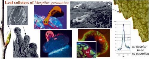
New data on the micromorphology, histochemistry, and fluorescence of colleters on leaf structures at different stages of development (leaf blade, stipules, and petiole) of Mespilus germanica L. are presented. Colleters are found on the tips of the teeth of both young and mature leaf blades and stipules, less often on the petioles. The leaf veins approach the leaf tooth, but no vascularization was found in the colleter. On leaf structures inside the bud, young colleters were observed in the form of finger-shaped or rounded outgrowths consisting of isodiametric cells. Mature colleters are multicellular secretory structures that have a head on a short stalk. The central part of the head consists of densely packed parenchymal cells, which are surrounded by radially elongated palisade-like secretory cells covered with a cuticle. The main secretion process of the colleter falls on the period of active growth of leaf structures. The secreted substances accumulated in the intercellular spaces of the palisade-like cells of the head and then were released outside in the form of translucent vesicles. The secretion products were released when the cuticle was ruptured and spread over the surface of the head and tooth of the leaf blade and stipules. After the end of secretion, the sizes of the head of the colleter decreased, and an abscission zone appeared in the cells of the colleter stalk, along the border of which a fracture occurred when the head fell off. Histochemical analysis of the contents of the colleter showed the presence of polysaccharides, especially at a young age, substances of a phenolic nature and lipids at a more mature age. In the fluorescence spectrum of young leaf colleter secretion, a peak at 671–672 nm was observed upon excitation at 405 and 473 nm. The obtained data on Mespilus germanica L. colleter can be used in the taxonomy of Pyrinae and Rosaceae.
中文翻译:
Mespilus germanica L.(蔷薇科)叶子中的收集器:微形态学、组织化学和荧光
提出了德国棉铃草不同发育阶段(叶片、托叶和叶柄)叶结构收集器的微形态学、组织化学和荧光的新数据。收集器位于年轻和成熟叶片和托叶的齿尖上,较少见于叶柄上。叶脉接近叶齿,但收集器中未发现血管化。在芽内的叶子结构上,观察到幼小的收集器呈由等径细胞组成的指状或圆形的生长物形式。成熟的收集器是多细胞分泌结构,头部长在短柄上。头部的中央部分由密集的实质细胞组成,周围是径向拉长的栅栏状分泌细胞,上面覆盖着角质层。收集器的主要分泌过程落在叶结构的活跃生长时期。分泌的物质积聚在头部栅栏状细胞的细胞间隙中,然后以半透明囊泡的形式释放到外面。角质层破裂时分泌产物释放出来,遍布叶片和托叶的头和齿的表面。分泌结束后,收集器头部尺寸减小,收集器柄细胞出现脱落区,头部脱落时沿其边界发生断裂。对收集器内容物的组织化学分析显示,特别是在年轻时,存在多糖;在较成熟的年龄,存在酚类物质和脂质。在幼叶收集器分泌物的荧光光谱中,在 405 和 473 nm 激发时观察到 671-672 nm 处的峰值。获得的德国Mespilus germanica L. colleter 数据可用于梨亚科和蔷薇科的分类学。


 京公网安备 11010802027423号