Journal of Molecular Histology ( IF 3.2 ) Pub Date : 2023-09-20 , DOI: 10.1007/s10735-023-10153-6 Renfeng Xu 1, 2 , Siting Shen 2 , Defan Wang 3 , Jianqing Ye 1 , Shiting Song 2 , Zhengchao Wang 1 , Zhicao Yue 2
|
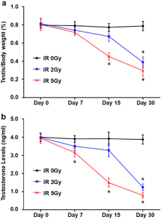
Testis, as a key organ for maintaining male fertility, are extremely sensitive to ionizing radiation (IR). IR-induced testicular dysfunction and infertility are common adverse effects of radiation therapy in patients with pelvic cancer. To study the phenotype and mechanism of IR-induced testicular injury, the mice were irradiated with different radiation doses (0, 2 and 5 Gy) below the semi-lethal dose for one month. Our present results showed that testicular weight and the serum testosterone levels significantly decreased with the structural injury of the testis in an IR dose-dependent manner, indicating that IR caused not only the structural damage of the testis, but also the functional damage. Further analysis by TUNEL staining and Western blotting found that IR induced the apoptosis in a dose-dependent manner as indicated by increased expressions of cleaved caspase3, p53 and Bax on Day 15 after IR treatment. Combined with significantly increased oxidative stress, these results indicated that IR-induced testicular damage may be a long-term, progressively aggravated process, accompanied by apoptosis. Given the role of autophagy in apoptosis, the present study also detected and analyzed the localization and expressions of autophagy-related proteins LC-3I/II, beclin1, p62 and Atg12 in testicular cells, and found that LC-3II, beclin1 and Atg12 expressions significantly increased in the testicular cells of mice irradiated with 2 Gy and 5 Gy, while p62 expression significantly decreased with 5 Gy, implying autophagy was involved in the apoptosis of testicular cells induced by IR. Furthermore, the expressions of HIF-1α and BNIP3 were significantly enhanced in the testis cells of mice irradiated with 2 Gy and 5 Gy, suggesting the potential role of HIF-1α/BNIP3-mediated autophagy in the apoptosis of testicular cells induced by IR. Taken together, our findings demonstrated that HIF-1α/BNIP3-mediated autophagy not only plays a protective effect on the testicular cells of mice irradiated with 2 Gy, but also induces the apoptosis of the testicular cells of mice irradiated with 5 Gy, indicating the double effects on apoptosis, which will help us further understanding the molecular mechanisms of IR-induced testicular injury, and will facilitate us further studies on testicular radioprotection.
中文翻译:
HIF-1α介导的自噬在电离辐射引起的睾丸损伤中的作用
睾丸作为维持男性生育能力的关键器官,对电离辐射(IR)极其敏感。红外线引起的睾丸功能障碍和不育是盆腔癌患者放射治疗的常见不良反应。为了研究红外线引起的睾丸损伤的表型和机制,对小鼠进行低于半致死剂量的不同辐射剂量(0、2和5 Gy)照射1个月。我们目前的结果表明,随着睾丸结构损伤,睾丸重量和血清睾酮水平显着下降,且呈IR剂量依赖性方式,表明IR不仅引起睾丸结构损伤,而且还引起功能损伤。通过 TUNEL 染色和蛋白质印迹进一步分析发现,IR 以剂量依赖性方式诱导细胞凋亡,如 IR 处理后第 15 天裂解的 caspase3、p53 和 Bax 的表达增加所表明的。结合显着增加的氧化应激,这些结果表明IR引起的睾丸损伤可能是一个长期的、逐渐加重的过程,并伴有细胞凋亡。鉴于自噬在细胞凋亡中的作用,本研究还检测并分析了自噬相关蛋白 LC-3I/II、beclin1、p62 和 Atg12 在睾丸细胞中的定位和表达情况,发现 LC-3II、beclin1 和 Atg12 表达2 Gy和5 Gy照射小鼠睾丸细胞中p62表达显着增加,而5 Gy照射后p62表达显着下降,表明自噬参与了IR诱导的睾丸细胞凋亡。此外,经2 Gy和5 Gy照射的小鼠睾丸细胞中HIF-1α和BNIP3的表达显着增强,表明HIF-1α/BNIP3介导的自噬在IR诱导的睾丸细胞凋亡中具有潜在作用。综上所述,我们的研究结果表明,HIF-1α/BNIP3介导的自噬不仅对2 Gy照射的小鼠睾丸细胞起到保护作用,而且还诱导5 Gy照射的小鼠睾丸细胞凋亡,这表明细胞凋亡的双重作用,将有助于我们进一步了解IR引起睾丸损伤的分子机制,并有利于我们对睾丸辐射防护的进一步研究。


 京公网安备 11010802027423号