Journal of Molecular Histology ( IF 3.2 ) Pub Date : 2023-09-27 , DOI: 10.1007/s10735-023-10160-7 Shitong Zhao 1 , Jingjing Cui 2 , Yuqing Wang 2 , Dongsheng Xu 2 , Yuxin Su 2 , Jie Ma 3 , Xuefeng Gong 1 , Wanzhu Bai 2 , Jia Wang 2 , Rui Cao 1
|
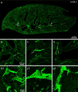
In order to demonstrate the intricate interconnection of pulmonary lymphatic vessels, blood vessels, and nerve fibers, the rat lung was selected as the target and sliced at the thickness of 100 μm for multiply immunofluorescence staining with lymphatic vessel endothelial hyaluronan receptor 1 (LYVE-1), alpha smooth muscle actin (α-SMA), phalloidin, cluster of differentiation 31 (CD31), and protein gene product 9.5 (PGP9.5) antibodies. Taking the advantages of the thicker tissue section and confocal microscopy, the labeled pulmonary lymphatic vessels, blood vessels, and nerve fibers were demonstrated in rather longer distance, which was more convenient to reconstruct a three-dimensional (3D) view for analyzing their spatial correlation in detail. It was clear that LYVE-1+ lymphatic vessels were widely distributed in pulmonary lobules and closely to the lobar bronchus. Through 3D reconstruction, it was also demonstrated that LYVE-1+ lymphatic vessels ran parallel to or around the α-SMA+ venules, phalloidin+ arterioles and CD31+ capillaries, with PGP9.5+ nerve fibers traversing alongside or wrapping around them, forming a lymphatic, vascular and neural network in the lung. By this study, we provide a detailed histological view to highlight the spatial correlation of pulmonary lymphatic, vascular and neural network, which may help us for insight into the functional role of this network under the physiological and pathological conditions.
中文翻译:
通过共聚焦显微镜对大鼠肺中淋巴管、血管和神经网络进行三维可视化
为了证明肺淋巴管、血管和神经纤维之间错综复杂的相互联系,选择大鼠肺作为靶标,切成100μm厚的切片,用淋巴管内皮透明质酸受体1(LYVE-1)进行多重免疫荧光染色。 )、α 平滑肌肌动蛋白 (α-SMA)、鬼笔环肽、分化簇 31 (CD31) 和蛋白质基因产物 9.5 (PGP9.5) 抗体。利用较厚的组织切片和共焦显微镜的优点,标记的肺淋巴管、血管和神经纤维在相当远的距离上得到展示,这更方便重建三维(3D)视图以分析它们的空间相关性详细。很明显,LYVE-1 +淋巴管广泛分布于肺小叶,且靠近叶支气管。通过3D重建,还证明LYVE-1 +淋巴管平行于或围绕α-SMA +小静脉、鬼笔环肽+小动脉和CD31 +毛细血管,PGP9.5 +神经纤维沿着或缠绕在它们周围,形成肺部的淋巴管、血管和神经网络。通过这项研究,我们提供了详细的组织学视图,以突出肺淋巴管、血管和神经网络的空间相关性,这可能有助于我们深入了解该网络在生理和病理条件下的功能作用。


 京公网安备 11010802027423号