Micron ( IF 2.4 ) Pub Date : 2023-10-01 , DOI: 10.1016/j.micron.2023.103547 Rutika Sansaria 1 , Krishanu Dey Das 1 , Alwin Poulose 2
|
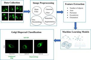
The Golgi body is a critical organelle in eukaryotic cells responsible for processing and modifying proteins and lipids. Under certain conditions, such as stress, disease, or ageing, the Golgi structure alters. Therefore, understanding the mechanisms that regulate Golgi dispersion has significant research contributions to identifying disease. However, there is a lack of tools to quantify the Golgi dispersion datasets. In this paper, we aim to automate the process of quantification of Golgi dispersion and use extracted features to classify dispersed Golgi images from undispersed Golgi images using machine learning models. First, we collected confocal microscopy images of transiently transfected HeLa cells expressing Galactose-1-phosphate uridylyltransferase (GALT)- green fluorescent protein (GFP) to quantify Golgi dispersal and classification. For the quantification, we introduced automated image processing and segmentation by applying mean and Gaussian filters. Then we used Otsu thresholding on preprocessed images and watershed segmentation to refine the segmentation of dispersed Golgi particles. In the case of classification, we extracted features from the Golgi dispersal images and classified them into empty vector (EV) versus CARP1 ring mutant (CARP1 RM) and empty vector (EV) versus CARP1 wildtype (CARP1 WT) classes. Our approach used machine-learning models, including logistic regression, decision tree, random forest, Naive Bayes, k-Nearest Neighbor (KNN), and gradient boosting for dispersed Golgi image classification. The experiment results show that our quantification technique on Golgi dispersal images reached 65% classification accuracy when the system uses a gradient boosting classifier for EV vs. CARP1 WT classification. Furthermore, our approach achieved 65% classification accuracy using a random forest classifier for EV vs. CARP1 RM classification.
中文翻译:
使用机器学习模型对高尔基体分散和分类进行量化
高尔基体是真核细胞中负责加工和修饰蛋白质和脂质的关键细胞器。在某些条件下,例如压力、疾病或衰老,高尔基体结构会发生变化。因此,了解调节高尔基体分散的机制对于识别疾病具有重要的研究贡献。然而,缺乏量化高尔基体离散数据集的工具。在本文中,我们的目标是自动化高尔基体分散的量化过程,并使用提取的特征使用机器学习模型将分散的高尔基体图像与未分散的高尔基体图像进行分类。首先,我们收集了表达半乳糖-1-磷酸尿苷酰转移酶(GALT)-绿色荧光蛋白(GFP)的瞬时转染的 HeLa 细胞的共聚焦显微镜图像,以量化高尔基体的分散和分类。为了进行量化,我们通过应用均值滤波器和高斯滤波器引入了自动图像处理和分割。然后,我们对预处理图像使用 Otsu 阈值处理和分水岭分割来细化分散高尔基体颗粒的分割。在分类的情况下,我们从高尔基体分散图像中提取特征,并将其分类为空矢量(EV)与CARP1环突变体(CARP1 RM)和空矢量(EV)与CARP1野生型(CARP1 WT)类。我们的方法使用机器学习模型,包括逻辑回归、决策树、随机森林、朴素贝叶斯、k 最近邻 (KNN) 和用于分散高尔基体图像分类的梯度提升。实验结果表明,当系统使用梯度增强分类器进行 EV 与 CARP1 WT 分类时,我们对高尔基体分散图像的量化技术达到了 65% 的分类精度。此外,我们的方法使用随机森林分类器进行 EV 与 CARP1 RM 分类,实现了 65% 的分类准确率。


 京公网安备 11010802027423号