当前位置:
X-MOL 学术
›
Genes Cells
›
论文详情
Our official English website, www.x-mol.net, welcomes your feedback! (Note: you will need to create a separate account there.)
Dynamics of actomyosin filaments in the contractile ring revealed by ultrastructural analysis
Genes to Cells ( IF 2.1 ) Pub Date : 2023-10-16 , DOI: 10.1111/gtc.13073 Takeru Arima 1 , Keisuke Okita 1 , Shigehiko Yumura 1
Genes to Cells ( IF 2.1 ) Pub Date : 2023-10-16 , DOI: 10.1111/gtc.13073 Takeru Arima 1 , Keisuke Okita 1 , Shigehiko Yumura 1
Affiliation
|
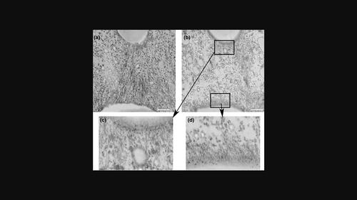
Cytokinesis, the final process of cell division, involves the accumulation of actin and myosin II filaments at the cell's equator, forming a contractile ring that facilitates the division into two daughter cells. While light microscopy has provided valuable insights into the molecular mechanism of this process, it has limitations in examining individual filaments in vivo. In this study, we utilized transmission electron microscopy to observe actin and myosin II filaments in the contractile rings of dividing Dictyostelium cells. To synchronize cytokinesis, we developed a novel method that allowed us to visualize dividing cells undergoing cytokinesis with a frequency as high as 18%. This improvement enabled us to examine the lengths and alignments of individual filaments within the contractile rings. As the furrow constricted, the length of actin filaments gradually decreased. Moreover, both actin and myosin II filaments reoriented perpendicularly to the long axis during furrow constriction. Through experiments involving myosin II null cells, we discovered that myosin II plays a role in regulating both the lengths and alignments of actin filaments. Additionally, dynamin-like protein A was found to contribute to regulating the length of actin filaments, while cortexillins were involved in regulating their alignment.
中文翻译:

超微结构分析揭示收缩环中肌动球蛋白丝的动力学
细胞分裂是细胞分裂的最后过程,涉及肌动蛋白和肌球蛋白 II 丝在细胞赤道处的积累,形成一个收缩环,促进分裂成两个子细胞。虽然光学显微镜为这一过程的分子机制提供了有价值的见解,但它在体内检查单个细丝方面存在局限性。在这项研究中,我们利用透射电子显微镜观察分裂的盘基网柄菌细胞收缩环中的肌动蛋白和肌球蛋白 II 丝。为了同步胞质分裂,我们开发了一种新方法,使我们能够可视化正在进行胞质分裂的分裂细胞,频率高达 18%。这一改进使我们能够检查收缩环内单个细丝的长度和排列。随着沟槽收缩,肌动蛋白丝的长度逐渐缩短。此外,在沟收缩期间,肌动蛋白和肌球蛋白 II 丝都垂直于长轴重新定向。通过涉及肌球蛋白 II 无效细胞的实验,我们发现肌球蛋白 II 在调节肌动蛋白丝的长度和排列方面发挥着作用。此外,动力样蛋白 A 被发现有助于调节肌动蛋白丝的长度,而皮质西林则参与调节其排列。
更新日期:2023-10-16
中文翻译:
超微结构分析揭示收缩环中肌动球蛋白丝的动力学
细胞分裂是细胞分裂的最后过程,涉及肌动蛋白和肌球蛋白 II 丝在细胞赤道处的积累,形成一个收缩环,促进分裂成两个子细胞。虽然光学显微镜为这一过程的分子机制提供了有价值的见解,但它在体内检查单个细丝方面存在局限性。在这项研究中,我们利用透射电子显微镜观察分裂的盘基网柄菌细胞收缩环中的肌动蛋白和肌球蛋白 II 丝。为了同步胞质分裂,我们开发了一种新方法,使我们能够可视化正在进行胞质分裂的分裂细胞,频率高达 18%。这一改进使我们能够检查收缩环内单个细丝的长度和排列。随着沟槽收缩,肌动蛋白丝的长度逐渐缩短。此外,在沟收缩期间,肌动蛋白和肌球蛋白 II 丝都垂直于长轴重新定向。通过涉及肌球蛋白 II 无效细胞的实验,我们发现肌球蛋白 II 在调节肌动蛋白丝的长度和排列方面发挥着作用。此外,动力样蛋白 A 被发现有助于调节肌动蛋白丝的长度,而皮质西林则参与调节其排列。


 京公网安备 11010802027423号