Calcified Tissue International ( IF 4.2 ) Pub Date : 2023-11-01 , DOI: 10.1007/s00223-023-01149-1 Quirina C B S Thio 1, 2, 3 , Olivier D R van Wulfften Palthe 1, 2 , Jos A M Bramer 1 , Thomas F DeLaney 4 , Miriam A Bredella 5 , David W Dempster 6, 7 , Hua Zhou 7 , Francis J Hornicek 8 , Yen-Lin E Chen 4 , Joseph H Schwab 2
|
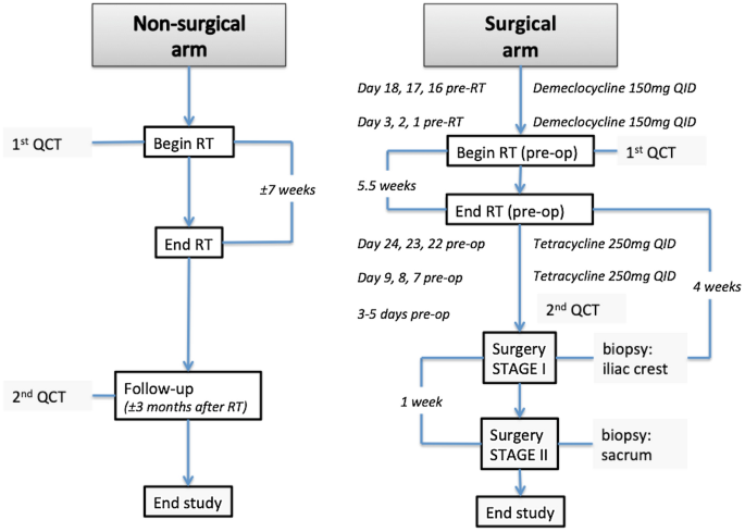
Despite the risk of complications, high dose radiation therapy is increasingly utilized in the management of selected bone malignancies. In this study, we investigate the impact of moderate to high dose radiation (over 50 Gy) on bone metabolism and structure. Between 2015 and 2018, patients with a primary malignant bone tumor of the sacrum that were either treated with high dose definitive radiation only or a combination of moderate to high dose radiation and surgery were prospectively enrolled at a single institution. Quantitative CTs were performed before and after radiation to determine changes in volumetric bone mineral density (BMD) of the irradiated and non-irradiated spine. Bone histomorphometry was performed on biopsies of the irradiated sacrum and the non-irradiated iliac crest of surgical patients using a quadruple tetracycline labeling protocol. In total, 9 patients were enrolled. Two patients received radiation only (median dose 78.3 Gy) and 7 patients received a combination of preoperative radiation (median dose 50.4 Gy), followed by surgery. Volumetric BMD of the non-irradiated lumbar spine did not change significantly after radiation, while the BMD of the irradiated sacrum did (pre-radiation median: 108.0 mg/cm3 (IQR 91.8–167.1); post-radiation median: 75.3 mg/cm3 (IQR 57.1–110.2); p = 0.010). The cancellous bone of the non-irradiated iliac crest had a stable bone formation rate, while the irradiated sacrum showed a significant decrease in bone formation rate [pre-radiation median: 0.005 mm3/mm2/year (IQR 0.003–0.009), post-radiation median: 0.001 mm3/mm2/year (IQR 0.001–0.001); p = 0.043]. Similar effects were seen in the cancellous and endocortical envelopes. This pilot study shows a decrease of volumetric BMD and bone formation rate after high-dose radiation therapy. Further studies with larger cohorts and other endpoints are needed to get more insight into the effect of radiation on bone. Level of evidence: IV.
中文翻译:
初步研究:放射治疗对骨矿物质密度和骨代谢的短期影响
尽管存在并发症的风险,高剂量放射治疗越来越多地用于治疗选定的骨恶性肿瘤。在这项研究中,我们研究了中高剂量辐射(超过 50 Gy)对骨代谢和结构的影响。2015 年至 2018 年间,单一机构前瞻性招募了原发性骶骨恶性骨肿瘤患者,这些患者要么仅接受高剂量确定性放射治疗,要么接受中高剂量放射和手术相结合的治疗。在放射前后进行定量 CT 检查,以确定受照射和未受照射脊柱的体积骨矿物质密度 (BMD) 的变化。使用四环素标记方案对手术患者的受辐射骶骨和未受辐射的髂嵴活检进行骨组织形态测定。总共有 9 名患者入组。两名患者仅接受放疗(中位剂量 78.3 Gy),7 名患者接受术前联合放疗(中位剂量 50.4 Gy),然后进行手术。未照射腰椎的体积 BMD 在照射后没有显着变化,而照射后骶骨的 BMD 却发生了显着变化(照射前中位数:108.0 mg/cm 3 (IQR 91.8–167.1);照射后中位数:75.3 mg/cm 3 cm 3 (IQR 57.1–110.2);p = 0.010)。未辐照的髂嵴松质骨具有稳定的骨形成率,而辐照后的骶骨骨形成率显着下降[辐照前中位数:0.005 mm 3 /mm 2 /年(IQR 0.003–0.009),辐射后中位数:0.001 mm 3 /mm 2 /年(IQR 0.001–0.001);p = 0.043]。在松质纤维和皮质内包膜中也观察到类似的效果。这项初步研究表明,高剂量放射治疗后体积骨密度和骨形成率有所下降。需要对更大的队列和其他终点进行进一步的研究,以更深入地了解辐射对骨骼的影响。证据级别:IV。


 京公网安备 11010802027423号