当前位置:
X-MOL 学术
›
Comput. Animat. Virtual Worlds
›
论文详情
Our official English website, www.x-mol.net, welcomes your feedback! (Note: you will need to create a separate account there.)
A 3D visualization-based augmented reality application for brain tumor segmentation
Computer Animation and Virtual Worlds ( IF 1.1 ) Pub Date : 2023-11-03 , DOI: 10.1002/cav.2223 Mohamed Amine Guerroudji 1 , Kahina Amara 1 , Mohamed Lichouri 2 , Nadia Zenati 1 , Mostefa Masmoudi 1
Computer Animation and Virtual Worlds ( IF 1.1 ) Pub Date : 2023-11-03 , DOI: 10.1002/cav.2223 Mohamed Amine Guerroudji 1 , Kahina Amara 1 , Mohamed Lichouri 2 , Nadia Zenati 1 , Mostefa Masmoudi 1
Affiliation
|
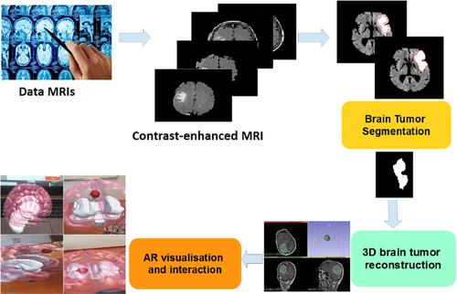
Every year on June 8th, the globe observes World Brain Tumor Day to raise awareness and educate people about brain cancer, encompassing both noncancerous (benign) and cancerous (malignant) growths. Research in the field of brain cancer plays a vital role in supporting medical professionals. In this context, augmented reality (AR) technology has emerged as a valuable tool, enabling surgeons to visualize underlying structures and offering a cost-effective and time-efficient alternative. Our study focuses on the efficient segmentation of brain tumor classes using Magnetic Resonance Imaging (MRI) and incorporates a three-stage approach: preprocessing, segmentation, and 3D reconstruction & AR display. In the preprocessing stage, a Gaussian filter is applied to mitigate intensity heterogeneity. Segmentation and detection are achieved using active geometric contour models, complemented by morphological operations. To establish 3D brain tumor reconstruction, a genuine scene is virtually integrated using 3D Slicer software. The proposed methodology was validated using a genuine patient dataset comprising 496 MRI scans obtained from the local Bab El Oued university hospital center. The results demonstrate the effectiveness of our approach in achieving accurate 3D brain tumor reconstruction, efficient tumor extraction, and augmented reality visualization. The obtained segmentation results showcased an impressive accuracy of 98.61%, outperforming existing state-of-the-art methods and affirming the efficacy of our proposed strategy.
中文翻译:

基于 3D 可视化的增强现实应用程序用于脑肿瘤分割
每年 6 月 8 日,全球都会庆祝世界脑肿瘤日,以提高人们对脑癌(包括非癌性(良性)和癌性(恶性)生长)的认识并进行教育。脑癌领域的研究在支持医疗专业人员方面发挥着至关重要的作用。在这种背景下,增强现实 (AR) 技术已成为一种有价值的工具,使外科医生能够可视化底层结构,并提供一种经济有效且省时的替代方案。我们的研究重点是使用磁共振成像 (MRI) 对脑肿瘤类别进行有效分割,并采用三阶段方法:预处理、分割以及 3D 重建和 AR 显示。在预处理阶段,应用高斯滤波器来减轻强度异质性。分割和检测是使用主动几何轮廓模型实现的,并辅以形态学操作。为了建立 3D 脑肿瘤重建,需要使用 3D Slicer 软件对真实场景进行虚拟集成。所提出的方法使用真实的患者数据集进行了验证,该数据集包含从当地 Bab El Oued 大学医院中心获得的 496 幅 MRI 扫描图像。结果证明了我们的方法在实现精确 3D 脑肿瘤重建、高效肿瘤提取和增强现实可视化方面的有效性。获得的分割结果显示出高达 98.61% 的准确率,优于现有的最先进方法,并肯定了我们提出的策略的有效性。
更新日期:2023-11-03
中文翻译:
基于 3D 可视化的增强现实应用程序用于脑肿瘤分割
每年 6 月 8 日,全球都会庆祝世界脑肿瘤日,以提高人们对脑癌(包括非癌性(良性)和癌性(恶性)生长)的认识并进行教育。脑癌领域的研究在支持医疗专业人员方面发挥着至关重要的作用。在这种背景下,增强现实 (AR) 技术已成为一种有价值的工具,使外科医生能够可视化底层结构,并提供一种经济有效且省时的替代方案。我们的研究重点是使用磁共振成像 (MRI) 对脑肿瘤类别进行有效分割,并采用三阶段方法:预处理、分割以及 3D 重建和 AR 显示。在预处理阶段,应用高斯滤波器来减轻强度异质性。分割和检测是使用主动几何轮廓模型实现的,并辅以形态学操作。为了建立 3D 脑肿瘤重建,需要使用 3D Slicer 软件对真实场景进行虚拟集成。所提出的方法使用真实的患者数据集进行了验证,该数据集包含从当地 Bab El Oued 大学医院中心获得的 496 幅 MRI 扫描图像。结果证明了我们的方法在实现精确 3D 脑肿瘤重建、高效肿瘤提取和增强现实可视化方面的有效性。获得的分割结果显示出高达 98.61% 的准确率,优于现有的最先进方法,并肯定了我们提出的策略的有效性。


 京公网安备 11010802027423号