Journal of Molecular and Cellular Cardiology ( IF 5 ) Pub Date : 2023-11-09 , DOI: 10.1016/j.yjmcc.2023.10.005 Venkatesh Mallikarjun 1 , Bocheng Yin 2 , Laura R Caggiano 1 , Sydney Blimbaum 2 , Caitlin M Pavelec 1 , Jeffrey W Holmes 3 , Sarah E Ewald 2
|
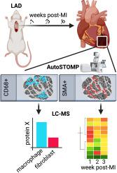
Myocardial infarction (MI) results from occlusion of blood supply to the heart muscle causing death of cardiac muscle cells. Following myocardial infarction (MI), extracellular matrix deposition and scar formation mechanically stabilize the injured heart as damaged myocytes undergo necrosis and removal. Fibroblasts and macrophages are key drivers of post-MI scar formation, maturation, and ongoing long-term remodelling; however, their individual contributions are difficult to assess from bulk analyses of infarct scar. Here, we employ state-of-the-art automated spatially targeted optical micro proteomics (autoSTOMP) to photochemically tag and isolate proteomes associated with subpopulations of fibroblasts (SMA+) and macrophages (CD68+) in the context of the native, MI tissue environment. Over a time course of 6-weeks post-MI, we captured dynamic changes in the whole-infarct proteome and determined that some of these protein composition signatures were differentially localized near SMA+ fibroblasts or CD68+ macrophages within the scar region. These results link specific cell populations to within-infarct protein remodelling and illustrate the distinct metabolic and structural processes underlying the observed physiology of each cell type.
中文翻译:
自动空间靶向光学微蛋白质组学识别大鼠心肌梗死疤痕成熟的成纤维细胞和巨噬细胞特异性调节
心肌梗塞 (MI) 是由于心肌血液供应阻塞导致心肌细胞死亡所致。心肌梗塞(MI)后,随着受损的心肌细胞坏死和去除,细胞外基质沉积和疤痕形成机械地稳定受损的心脏。成纤维细胞和巨噬细胞是心肌梗死后疤痕形成、成熟和持续长期重塑的关键驱动因素;然而,通过对梗塞疤痕的批量分析很难评估它们各自的贡献。在这里,我们采用最先进的自动化空间靶向光学微蛋白质组学 (autoSTOMP) 来光化学标记和分离与天然 MI 组织中的成纤维细胞 (SMA + ) 和巨噬细胞 (CD68 + ) 亚群相关的蛋白质组环境。在 MI 后 6 周的时间过程中,我们捕获了整个梗死蛋白质组的动态变化,并确定其中一些蛋白质组成特征差异性地定位在疤痕区域内的SMA +成纤维细胞或 CD68 +巨噬细胞附近。这些结果将特定细胞群与梗塞内蛋白质重塑联系起来,并说明了观察到的每种细胞类型的生理学背后的不同代谢和结构过程。


 京公网安备 11010802027423号