Forensic Science, Medicine and Pathology ( IF 1.8 ) Pub Date : 2023-11-16 , DOI: 10.1007/s12024-023-00751-x Victor Ibanez , Dario Jucker , Lars C. Ebert , Sabine Franckenberg , Akos Dobay
|
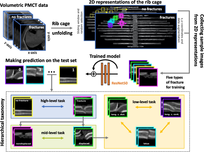
Human or time resources can sometimes fall short in medical image diagnostics, and analyzing images in full detail can be a challenging task. With recent advances in artificial intelligence, an increasing number of systems have been developed to assist clinicians in their work. In this study, the objective was to train a model that can distinguish between various fracture types on different levels of hierarchical taxonomy and detect them on 2D-image representations of volumetric postmortem computed tomography (PMCT) data. We used a deep learning model based on the ResNet50 architecture that was pretrained on ImageNet data, and we used transfer learning to fine-tune it to our specific task. We trained our model to distinguish between “displaced,” “nondisplaced,” “ad latus,” “ad longitudinem cum contractione,” and “ad longitudinem cum distractione” fractures. Radiographs with no fractures were correctly predicted in 95–99% of cases. Nondisplaced fractures were correctly predicted in 80–86% of cases. Displaced fractures of the “ad latus” type were correctly predicted in 17–18% of cases. The other two displaced types of fractures, “ad longitudinem cum contractione” and “ad longitudinem cum distractione,” were correctly predicted in 70–75% and 64–75% of cases, respectively. The model achieved the best performance when the level of hierarchical taxonomy was high, while it had more difficulties when the level of hierarchical taxonomy was lower. Overall, deep learning techniques constitute a reliable solution for forensic pathologists and medical practitioners seeking to reduce workload.
中文翻译:
使用深度学习根据死后计算机断层扫描图像对肋骨骨折类型进行分类
医学图像诊断中的人力或时间资源有时会不足,并且全面详细地分析图像可能是一项具有挑战性的任务。随着人工智能的最新进展,越来越多的系统被开发出来来协助临床医生的工作。在本研究中,目标是训练一个模型,该模型可以区分不同级别的分层分类中的各种骨折类型,并在体积死后计算机断层扫描 (PMCT) 数据的二维图像表示上检测它们。我们使用了基于 ResNet50 架构的深度学习模型,该模型在 ImageNet 数据上进行了预训练,并使用迁移学习对其进行微调以适应我们的特定任务。我们训练模型来区分“移位”、“非移位”、“横向”、“纵向结合收缩”和“纵向结合分散”骨折。95-99% 的病例正确预测了 X 光片上没有骨折的情况。80-86% 的病例正确预测无移位骨折。17-18% 的病例被正确预测为“ad latus”型移位骨折。另外两种移位类型的骨折,“纵向加收缩”和“纵向加牵引”,分别在 70-75% 和 64-75% 的病例中被正确预测。当层次分类水平较高时,该模型取得了最好的性能,而当层次分类水平较低时,该模型的性能较差。总体而言,深度学习技术为法医病理学家和医疗从业者寻求减少工作量提供了可靠的解决方案。


 京公网安备 11010802027423号