Journal of Electrocardiology ( IF 1.3 ) Pub Date : 2023-11-19 , DOI: 10.1016/j.jelectrocard.2023.11.007 Juan M Farina 1 , Thanaboon Yinadsawaphan 2 , Dawn E Jaroszewski 3 , Mohamed R Aly 3 , Michael Botros 3 , Kamal P Cheema 2 , Olubadewa A Fatunde 2 , Dan Sorajja 2
|
Background
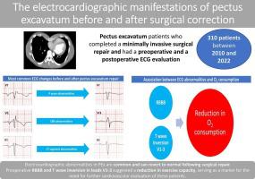
Pectus excavatum (PEx) can cause cardiopulmonary limitations due to cardiac compression and displacement. There is limited data on electrocardiogram (ECG) alterations before and after PEx surgical repair, and ECG findings suggesting cardiopulmonary limitations have not been reported. The aim of this study is to explore ECG manifestations of PEx before and after surgery including associations with exercise capacity.
Methods
A retrospective review of PEx patients who underwent primary repair was performed. ECGs before and after surgical correction were evaluated and the associations between preoperative ECG abnormalities and cardiopulmonary function were investigated.
Results
In total, 310 patients were included (mean age 35.1 ± 11.6 years). Preoperative ECG findings included a predominant negative P wave morphology in V1, and this abnormal pattern significantly decreased from 86.9% to 57.4% (p < 0.001) postoperatively. The presence of abnormal P wave amplitude in lead II (>2.5 mm) significantly decreased from 7.1% to 1.6% postoperatively (p < 0.001). Right bundle branch block (RBBB) (9.4% versus 3.9%, p < 0.001), rsr' patterns (40.6% versus 12.9%, p < 0.001), and T wave inversion in leads V1-V3 (62.3% vs 37.7%, p < 0.001) were observed less frequently after surgery. Preoperative presence of RBBB (OR = 4.8; 95%CI 1.1–21.6) and T wave inversion in leads V1–3 (OR = 2.3; 95%CI 1.3–4.2) were associated with abnormal results in cardiopulmonary exercise testings.
Conclusion
Electrocardiographic abnormalities in PEx are frequent and can revert to normal following surgery. Preoperative RBBB and T wave inversion in leads V1–3 suggested a reduction in exercise capacity, serving as a marker for the need for further cardiovascular evaluation of these patients.
中文翻译:
漏斗胸矫正手术前后心电图表现
背景
漏斗胸 (PEx) 可因心脏受压和移位而导致心肺受限。关于 PEx 手术修复前后心电图 (ECG) 变化的数据有限,并且尚未报道表明心肺功能受限的心电图结果。本研究的目的是探讨 PEx 手术前后的心电图表现,包括与运动能力的关系。
方法
对接受初次修复的 PEx 患者进行了回顾性审查。评估手术矫正前后的心电图,并研究术前心电图异常与心肺功能之间的关联。
结果
总共包括 310 名患者(平均年龄 35.1 ± 11.6 岁)。术前心电图结果包括 V1 中主要为负性 P 波形态,术后这种异常形态从 86.9% 显着下降至 57.4% ( p < 0.001)。术后 II 导联异常 P 波振幅(>2.5 mm)从 7.1% 显着下降至 1.6%(p < 0.001)。右束支传导阻滞 (RBBB)(9.4% 对比 3.9%,p < 0.001)、rsr' 模式(40.6% 对比 12.9%,p < 0.001)以及 V1-V3 导联 T 波倒置(62.3% 对比 37.7%, p < 0.001)在手术后观察到的频率较低。术前存在 RBBB(OR = 4.8;95%CI 1.1-21.6)和 V1-3 导联 T 波倒置(OR = 2.3;95%CI 1.3-4.2)与心肺运动试验的异常结果相关。
结论
PEx 心电图异常很常见,并且可以在手术后恢复正常。术前 V1-3 导联的 RBBB 和 T 波倒置表明运动能力下降,作为需要对这些患者进行进一步心血管评估的标志。


 京公网安备 11010802027423号