Cell and Tissue Research ( IF 3.6 ) Pub Date : 2023-11-20 , DOI: 10.1007/s00441-023-03842-x Maurizio Mazzoni 1 , Luis Cabanillas 2, 3 , Anna Costanzini 4 , Filippo Caremoli 2, 5 , Mulugeta Million 2, 6 , Muriel Larauche 2 , Paolo Clavenzani 1 , Roberto De Giorgio 4 , Catia Sternini 2, 3
|
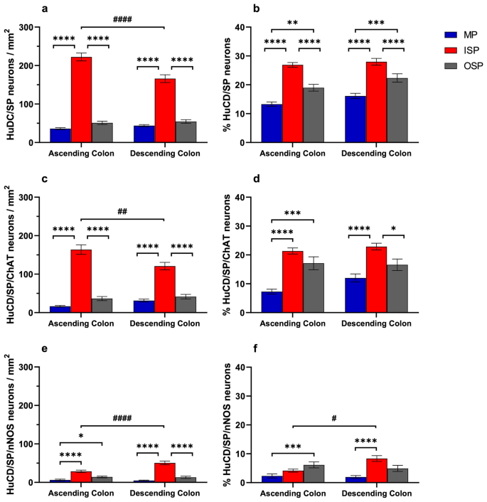
The pig is an important translational model for studying intestinal physiology and disorders for its many homologies with humans, including the organization of the enteric nervous system (ENS), the major regulator of gastrointestinal functions. This study focused on the quantification and neurochemical characterization of substance P (SP) neurons in the pig ascending (AC) and descending colon (DC) in wholemount preparations of the inner submucosal plexus (ISP), outer submucosal plexus (OSP), and myenteric plexus (MP). We used antibodies for the pan-neuronal marker HuCD, and choline acetyltransferase (ChAT) and neuronal nitric oxide synthase (nNOS), markers for excitatory and inhibitory transmitters, for multiple labeling immunofluorescence and high-resolution confocal microscopy. The highest density of SP immunoreactive (IR) neurons was in the ISP (222/mm2 in the AC, 166/mm2 in the DC), where they make up about a third of HuCD-IR neurons, compared to the OSP and MP (19–22% and 13–17%, respectively, P < 0.001–0.0001). HuCD/SP/ChAT-IR neurons (up to 23%) were overall more abundant than HuCD/SP/nNOS-IR neurons (< 10%). Most SP-IR neurons contained ChAT-IR (62–85%), whereas 18–38% contained nNOS-IR with the highest peak in the OSP. A subpopulation of SP-IR neurons contains both ChAT- and nNOS-IR with the highest peak in the OSP and ISP of DC (33–36%) and the lowest in the ISP of AC (< 10%, P < 0.001). SP-IR varicose fibers were abundant in the ganglia. This study shows that SP-IR neurons are functionally distinct with variable proportions in different plexuses in the AC and DC reflecting diverse functions of specific colonic regions.
中文翻译:
猪结肠粘膜下层和肌间丛中 P 物质肠神经元的分布、定量和表征
猪是研究肠道生理学和肠道疾病的重要转化模型,因为它与人类有许多相似之处,包括肠神经系统(ENS)的组织,这是胃肠道功能的主要调节器。本研究重点关注内粘膜下丛 (ISP)、外粘膜下丛 (OSP) 和肌间神经丛整体制备物中猪升结肠 (AC) 和降结肠 (DC) 中 P 物质 (SP) 神经元的定量和神经化学特征神经丛(MP)。我们使用泛神经元标记物 HuCD、胆碱乙酰转移酶 (ChAT) 和神经元一氧化氮合酶 (nNOS) 的抗体(兴奋性和抑制性递质的标记物)进行多重标记免疫荧光和高分辨率共聚焦显微镜。SP 免疫反应 (IR) 神经元密度最高的是 ISP(AC 中为222/mm 2 ,DC 中为 166/mm 2),与 OSP 和 OSP 相比,它们约占 HuCD-IR 神经元的三分之一。 MP(分别为 19–22% 和 13–17%,P < 0.001–0.0001)。HuCD/SP/ChAT-IR 神经元(高达 23%)总体上比 HuCD/SP/nNOS-IR 神经元(< 10%)更丰富。大多数 SP-IR 神经元含有 ChAT-IR (62-85%),而 18-38% 含有 nNOS-IR,在 OSP 中具有最高峰值。SP-IR 神经元亚群同时包含 ChAT- 和 nNOS-IR,DC 的 OSP 和 ISP 中峰值最高(33-36%),AC 的 ISP 中峰值最低(< 10%,P < 0.001)。SP-IR 曲张纤维在神经节中丰富。这项研究表明,AC 和 DC 不同丛中的 SP-IR 神经元功能不同,比例不同,反映了特定结肠区域的不同功能。


 京公网安备 11010802027423号