当前位置:
X-MOL 学术
›
J. Comp. Neurol.
›
论文详情
Our official English website, www.x-mol.net, welcomes your feedback! (Note: you will need to create a separate account there.)
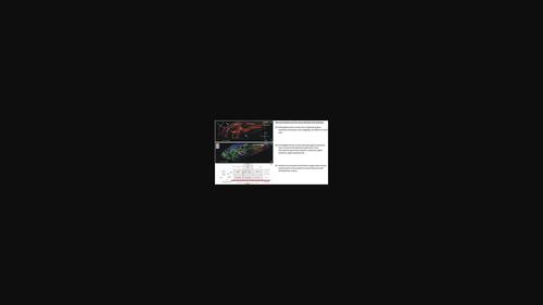
Genoarchitectonics of the larval zebrafish diencephalon
The Journal of Comparative Neurology ( IF 2.5 ) Pub Date : 2023-11-20 , DOI: 10.1002/cne.25549 Mario F Wullimann 1, 2 , Nouwar Mokayes 1 , Inbal Shainer 1 , Enrico Kuehn 1 , Herwig Baier 1
The Journal of Comparative Neurology ( IF 2.5 ) Pub Date : 2023-11-20 , DOI: 10.1002/cne.25549 Mario F Wullimann 1, 2 , Nouwar Mokayes 1 , Inbal Shainer 1 , Enrico Kuehn 1 , Herwig Baier 1
Affiliation
|
The brain is spatially organized into subdivisions, nuclei and areas, which often correspond to functional and developmental units. A segmentation of brain regions in the form of a consensus atlas facilitates mechanistic studies and is a prerequisite for sharing information among neuroanatomists. Gene expression patterns objectively delineate boundaries between brain regions and provide information about their developmental and evolutionary histories. To generate a detailed molecular map of the larval zebrafish diencephalon, we took advantage of the Max Planck Zebrafish Brain (mapzebrain) atlas, which aligns hundreds of transcript and transgene expression patterns in a shared coordinate system. Inspection and co-visualization of close to 50 marker genes have allowed us to resolve the tripartite prosomeric scaffold of the diencephalon at unprecedented resolution. This approach clarified the genoarchitectonic partitioning of the alar diencephalon into pretectum (alar part of prosomere P1), thalamus (alar part of prosomere P2, with habenula and pineal complex), and prethalamus (alar part of prosomere P3). We further identified the region of the nucleus of the medial longitudinal fasciculus, as well as the posterior and anterior parts of the posterior tuberculum, as molecularly distinct basal parts of prosomeres 1, 2, and 3, respectively. Some of the markers examined allowed us to locate glutamatergic, GABAergic, dopaminergic, serotoninergic, and various neuropeptidergic domains in the larval zebrafish diencephalon. Our molecular neuroanatomical approach has thus (1) yielded an objective and internally consistent interpretation of the prosomere boundaries within the zebrafish forebrain; has (2) produced a list of markers, which in sparse combinations label the subdivisions of the diencephalon; and is (3) setting the stage for further functional and developmental studies in this vertebrate brain.
中文翻译:

斑马鱼幼虫间脑的基因结构
大脑在空间上被组织成分区、核和区域,它们通常对应于功能和发育单位。以共识图谱形式对大脑区域进行分割有助于机制研究,也是神经解剖学家之间共享信息的先决条件。基因表达模式客观地描绘了大脑区域之间的界限,并提供有关其发育和进化历史的信息。为了生成斑马鱼幼虫间脑的详细分子图谱,我们利用了马克斯·普朗克斑马鱼大脑 (mapzebrain) 图集,该图集在共享坐标系中对齐了数百个转录本和转基因表达模式。对近 50 个标记基因的检查和联合可视化使我们能够以前所未有的分辨率解析间脑的三部分前体支架。这种方法阐明了翼间脑的基因结构划分为前体(前体 P1 的翼部)、丘脑(前体 P2 的翼部,具有缰核和松果体复合体)和丘脑前体(前体 P3 的翼部)。我们进一步确定了内侧纵束核区域以及后结节的后部和前部分别为前体 1、2 和 3 分子上不同的基底部分。检查的一些标记使我们能够在斑马鱼幼虫间脑中定位谷氨酸能、GABA能、多巴胺能、血清素能和各种神经肽能结构域。因此,我们的分子神经解剖学方法(1)对斑马鱼前脑内的前体边界产生了客观且内部一致的解释;(2) 生成了一系列标记,这些标记以稀疏组合标记间脑的细分;(3) 为脊椎动物大脑的进一步功能和发育研究奠定基础。
更新日期:2023-11-20
中文翻译:
斑马鱼幼虫间脑的基因结构
大脑在空间上被组织成分区、核和区域,它们通常对应于功能和发育单位。以共识图谱形式对大脑区域进行分割有助于机制研究,也是神经解剖学家之间共享信息的先决条件。基因表达模式客观地描绘了大脑区域之间的界限,并提供有关其发育和进化历史的信息。为了生成斑马鱼幼虫间脑的详细分子图谱,我们利用了马克斯·普朗克斑马鱼大脑 (mapzebrain) 图集,该图集在共享坐标系中对齐了数百个转录本和转基因表达模式。对近 50 个标记基因的检查和联合可视化使我们能够以前所未有的分辨率解析间脑的三部分前体支架。这种方法阐明了翼间脑的基因结构划分为前体(前体 P1 的翼部)、丘脑(前体 P2 的翼部,具有缰核和松果体复合体)和丘脑前体(前体 P3 的翼部)。我们进一步确定了内侧纵束核区域以及后结节的后部和前部分别为前体 1、2 和 3 分子上不同的基底部分。检查的一些标记使我们能够在斑马鱼幼虫间脑中定位谷氨酸能、GABA能、多巴胺能、血清素能和各种神经肽能结构域。因此,我们的分子神经解剖学方法(1)对斑马鱼前脑内的前体边界产生了客观且内部一致的解释;(2) 生成了一系列标记,这些标记以稀疏组合标记间脑的细分;(3) 为脊椎动物大脑的进一步功能和发育研究奠定基础。


 京公网安备 11010802027423号