当前位置:
X-MOL 学术
›
J. Extracell. Vesicles
›
论文详情
Our official English website, www.x-mol.net, welcomes your feedback! (Note: you will need to create a separate account there.)
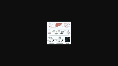
Three-dimensional reconstruction of interstitial extracellular vesicles in human liver as determined by electron tomography
Journal of Extracellular Vesicles ( IF 16.0 ) Pub Date : 2023-11-27 , DOI: 10.1002/jev2.12380 Roger Olofsson Bagge 1, 2 , Jens Berndtsson 3 , Ornella Urzì 1, 4 , Jan Lötvall 5 , Massimo Micaroni 3 , Rossella Crescitelli 1
Journal of Extracellular Vesicles ( IF 16.0 ) Pub Date : 2023-11-27 , DOI: 10.1002/jev2.12380 Roger Olofsson Bagge 1, 2 , Jens Berndtsson 3 , Ornella Urzì 1, 4 , Jan Lötvall 5 , Massimo Micaroni 3 , Rossella Crescitelli 1
Affiliation
|
Extracellular vesicles (EVs) are lipid bilayer nanoparticles involved in cell-cell communication that are released into the extracellular space by all cell types. The cargo of EVs includes proteins, lipids, nucleic acids, and metabolites reflecting their cell of origin. EVs have recently been isolated directly from solid tissues, and this may provide insights into how EVs mediate communication between cells in vivo. Even though EVs have been isolated from tissues, their point of origin when they are in the interstitial space has been uncertain. In this study, we performed three-dimensional (3D) reconstruction using transmission electron tomography of metastatic and normal liver tissues with a focus on the presence of EVs in the interstitium. After chemical fixation of the samples and subsequent embedding of tissue pieces in resin, ultrathin slices (300 nm) were cut and imaged on a 120 ekV transmission electron microscopy as a tilt series (a series of subsequent images tilted at different angles). These were then computationally illustrated in a 3D manner to reconstruct the imaged tissue volume. We identified the cells delimiting the interstitial space in both types of tissues, and small distinct spherical structures with a diameter of 30–200 nm were identified between the cells. These round structures appeared to be more abundant in metastatic tissue compared to normal tissue. We suggest that the observed spherical structures in the interstitium of the metastatic and non-metastatic liver represent EVs. This work thus provides the first 3D visualization of EVs in human tissue.
中文翻译:

电子断层扫描测定人肝脏间质细胞外囊泡的三维重建
细胞外囊泡 (EV) 是参与细胞间通讯的脂质双层纳米颗粒,所有细胞类型都会将其释放到细胞外空间。EV 的货物包括蛋白质、脂质、核酸和反映其细胞来源的代谢物。最近从实体组织中直接分离出 EV,这可能为了解 EV 如何介导体内细胞之间的通信提供见解。尽管细胞外囊泡已从组织中分离出来,但它们在间隙空间中的起源点仍不确定。在这项研究中,我们使用透射电子断层扫描对转移性和正常肝组织进行三维 (3D) 重建,重点关注间质中 EV 的存在。对样品进行化学固定并随后将组织块嵌入树脂中后,切割超薄切片(300 nm)并在 120 ekV 透射电子显微镜上作为倾斜系列成像(一系列以不同角度倾斜的后续图像)。然后以 3D 方式对这些进行计算说明,以重建成像的组织体积。我们确定了两种类型组织中界定间隙空间的细胞,并在细胞之间确定了直径为 30-200 nm 的小型独特球形结构。与正常组织相比,这些圆形结构在转移组织中似乎更丰富。我们认为转移性和非转移性肝脏间质中观察到的球形结构代表 EV。因此,这项工作首次实现了人体组织中 EV 的 3D 可视化。
更新日期:2023-11-28
中文翻译:
电子断层扫描测定人肝脏间质细胞外囊泡的三维重建
细胞外囊泡 (EV) 是参与细胞间通讯的脂质双层纳米颗粒,所有细胞类型都会将其释放到细胞外空间。EV 的货物包括蛋白质、脂质、核酸和反映其细胞来源的代谢物。最近从实体组织中直接分离出 EV,这可能为了解 EV 如何介导体内细胞之间的通信提供见解。尽管细胞外囊泡已从组织中分离出来,但它们在间隙空间中的起源点仍不确定。在这项研究中,我们使用透射电子断层扫描对转移性和正常肝组织进行三维 (3D) 重建,重点关注间质中 EV 的存在。对样品进行化学固定并随后将组织块嵌入树脂中后,切割超薄切片(300 nm)并在 120 ekV 透射电子显微镜上作为倾斜系列成像(一系列以不同角度倾斜的后续图像)。然后以 3D 方式对这些进行计算说明,以重建成像的组织体积。我们确定了两种类型组织中界定间隙空间的细胞,并在细胞之间确定了直径为 30-200 nm 的小型独特球形结构。与正常组织相比,这些圆形结构在转移组织中似乎更丰富。我们认为转移性和非转移性肝脏间质中观察到的球形结构代表 EV。因此,这项工作首次实现了人体组织中 EV 的 3D 可视化。


 京公网安备 11010802027423号