当前位置:
X-MOL 学术
›
J. Raman Spectrosc.
›
论文详情
Our official English website, www.x-mol.net, welcomes your feedback! (Note: you will need to create a separate account there.)
Feasibility of gold nanocones for collocated tip-enhanced Raman spectroscopy and atomic force microscope imaging
Journal of Raman Spectroscopy ( IF 2.5 ) Pub Date : 2023-11-30 , DOI: 10.1002/jrs.6625 Luke R. McCourt 1 , Ben S. Routley 1 , Michael G. Ruppert 2 , Andrew J. Fleming 1
Journal of Raman Spectroscopy ( IF 2.5 ) Pub Date : 2023-11-30 , DOI: 10.1002/jrs.6625 Luke R. McCourt 1 , Ben S. Routley 1 , Michael G. Ruppert 2 , Andrew J. Fleming 1
Affiliation
|
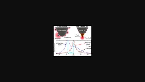
Microcantilever probes for tip-enhanced Raman spectroscopy (TERS) have a grainy metal coating that may exhibit multiple plasmon hotspots near the tip apex, which may compromise spatial resolution and introduce imaging artefacts. It is also possible that the optical hotspot may not occur at the mechanical apex, which introduces an offset between TERS and atomic force microscope maps. In this article, a gold nanocone TERS probe is designed and fabricated for 638 nm excitation. The imaging performance is compared to grainy probes by analysing high-resolution TERS cross-sections of single-walled carbon nanotubes. Compared to the tested conventional TERS probes, the nanocone probe exhibited a narrow spot diameter, comparable optical contrast, artefact-free images, and collocation of TERS and atomic force microscope topographic maps. The 1/
中文翻译:

金纳米锥用于并置尖端增强拉曼光谱和原子力显微镜成像的可行性
用于尖端增强拉曼光谱 (TERS) 的微悬臂梁探针具有颗粒状金属涂层,可能会在尖端顶点附近出现多个等离激元热点,这可能会损害空间分辨率并引入成像伪影。光学热点也可能不会出现在机械顶点,这会导致 TERS 和原子力显微镜图之间出现偏移。在本文中,设计并制造了用于 638 nm 激发的金纳米锥 TERS 探针。通过分析单壁碳纳米管的高分辨率 TERS 横截面,将成像性能与颗粒探针进行比较。与测试的传统 TERS 探针相比,纳米锥探针表现出窄光斑直径、可比的光学对比度、无伪影图像以及 TERS 和原子力显微镜地形图的搭配。1/
更新日期:2023-11-30
中文翻译:
金纳米锥用于并置尖端增强拉曼光谱和原子力显微镜成像的可行性
用于尖端增强拉曼光谱 (TERS) 的微悬臂梁探针具有颗粒状金属涂层,可能会在尖端顶点附近出现多个等离激元热点,这可能会损害空间分辨率并引入成像伪影。光学热点也可能不会出现在机械顶点,这会导致 TERS 和原子力显微镜图之间出现偏移。在本文中,设计并制造了用于 638 nm 激发的金纳米锥 TERS 探针。通过分析单壁碳纳米管的高分辨率 TERS 横截面,将成像性能与颗粒探针进行比较。与测试的传统 TERS 探针相比,纳米锥探针表现出窄光斑直径、可比的光学对比度、无伪影图像以及 TERS 和原子力显微镜地形图的搭配。1/


 京公网安备 11010802027423号