Calcified Tissue International ( IF 4.2 ) Pub Date : 2023-11-30 , DOI: 10.1007/s00223-023-01153-5 Pengbo Wang 1 , Xu Wang 1 , Hang Qian 1 , Jun Liu 1 , Gang Liu 2 , Ruisong Wang 3 , Ruiyu Liu 1
|
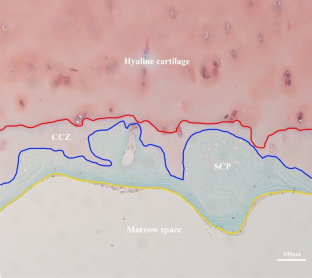
The study was aimed to investigate microarchitecture of osteochondral junction in patients with osteonecrosis of the femoral head (ONFH). We hypothesis that there were microarchitecture alternations in osteochondral junction and regional differences between the necrotic region (NR) and adjacent non-necrotic region(ANR) in patients with ONFH. Femoral heads with ONFH or femoral neck fracture were included in ONFH group (n = 11) and control group (n = 11). Cylindrical specimens were drilled on the NR/ANR of femoral heads in ONFH group and matched positions in control group (CO.NR/ CO.ANR). Histology, micro-CT, and scanning electron microscope were used to investigate microarchitecture of osteochondral junction. Layered analysis of subchondral bone plate was underwent. Mankin scores on NR were higher than that on ANR or CO.NR, respectively (P < 0.001, P < 0.001). Calcified cartilage zone on the NR and ANR was thinner than that on the CO.NR and CO.ANR, respectively (P = 0.002, P = 0.002). Tidemark roughness on the NR was larger than that on the ANR (P = 0.002). Subchondral bone plate of NR and ANR was thicker than that on the CON.NR and CON.ANR, respectively (P = 0.002, P = 0.009). Bone volume fraction of subchondral bone plate on the NR was significantly decreasing compared to ANR and CON.NR, respectively (P = 0.015, P = 0.002). Subchondral bone plate on the NR had larger area percentages and more numbers of micropores than ANR and CON.NR (P = 0.002/0.002, P = 0.002/0.002). Layered analysis showed that bone mass loss and hypomineralization were mainly on the cartilage side of subchondral bone plate in ONFH. There were microarchitecture alternations of osteochondral junction in ONFH, including thinned calcified cartilage zone, thickened subchondral bone plate, decreased bone mass, altered micropores, and hypomineralization of subchondral bone plate. Regional differences in microarchitecture of osteochondral junction were found between necrotic regions and adjacent non-necrotic regions. Subchondral bone plate in ONFH had uneven distribution of bone volume fraction and bone mineral density, which might aggravate cartilage degeneration by affecting the transmission of mechanical stresses.
中文翻译:
股骨头坏死患者骨软骨连接处微结构的改变
该研究旨在研究股骨头坏死(ONFH)患者骨软骨连接的微结构。我们假设ONFH患者的骨软骨连接处存在微结构改变,并且坏死区(NR)和邻近非坏死区(ANR)之间存在区域差异。合并ONFH或股骨颈骨折的股骨头分为ONFH组(n =11)和对照组(n =11)。ONFH组股骨头NR/ANR及对照组(CO.NR/CO.ANR)匹配位置钻取圆柱形标本。采用组织学、显微 CT 和扫描电子显微镜研究骨软骨连接的微结构。对软骨下骨板进行分层分析。NR 的 Mankin 评分分别高于 ANR 或 CO.NR(P < 0.001,P < 0.001)。NR 和 ANR 上的钙化软骨区分别比 CO.NR 和 CO.ANR 上的钙化软骨区薄(P = 0.002,P = 0.002)。NR 上的潮迹粗糙度大于 ANR 上的潮迹粗糙度(P = 0.002)。NR和ANR的软骨下骨板分别比CON.NR和CON.ANR的软骨下骨板厚(P =0.002,P =0.009)。与ANR和CON.NR相比,NR上软骨下骨板的骨体积分数分别显着降低(P = 0.015,P = 0.002)。NR上的软骨下骨板比ANR和CON.NR具有更大的面积百分比和更多的微孔数量(P = 0.002/0.002,P = 0.002/0.002)。分层分析显示ONFH的骨量丢失和矿化不足主要发生在软骨下骨板的软骨侧。ONFH中骨软骨连接处的微结构发生改变,包括钙化软骨区变薄、软骨下骨板增厚、骨量减少、微孔改变和软骨下骨板矿化低下。在坏死区域和相邻非坏死区域之间发现骨软骨连接微结构的区域差异。ONFH软骨下骨板骨体积分数和骨矿物质密度分布不均匀,可能通过影响机械应力的传递而加剧软骨退变。


 京公网安备 11010802027423号