Cell and Tissue Research ( IF 3.6 ) Pub Date : 2023-12-02 , DOI: 10.1007/s00441-023-03843-w Nadezhda V. Kalacheva , Talia T. Ginanova , Yaroslav O. Kamenev , Sergey I. Maslennikov , Igor Yu. Dolmatov
|
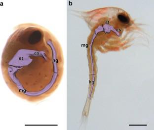
The digestive system structure in pre-zoea and zoea I larvae of the red king crab Paralithodes camtschaticus has been examined. During this development period, the digestive system consists of an esophagus, a stomach, a midgut (where the hepatopancreas ducts open), and a hindgut. The esophagus begins from the oral slit on the animal’s ventral side and extends vertically up to the junction with the cardiac stomach. The latter is followed by the pyloric stomach. At the stages under study, crabs have a cardiac-pyloric valve and a pyloric filter in the stomach already developed. The midgut begins with an expansion in the cephalothorax, enters the pleon, grows narrower there, and extends to somite 3 of pleon. The hepatopancreas is represented by a symmetrical paired gland which occupies almost the entire cephalothorax space and opens with its ducts at the junction of the pyloric stomach with the midgut. The hepatopancreas is divided into the anterior and posterior lobes. At the pre-zoea stage, the anterior lobes are large and filled with yolk. At the zoea I stage, the anterior lobes are smaller relative to the entire hepatopancreas, and the posterior lobes increase and form tubular outgrowths. It has been shown that during the transition from pre-zoea to zoea I, the number of mitochondria in enterocytes increases and a peritrophic membrane forms in the midgut. These changes are probably associated with the transition to independent living and feeding.
中文翻译:
红帝王蟹 Paralithodes camtschaticus 的前幼虫和幼虫 I 期幼虫消化系统的形态和超微结构 (Tilesius, 1815)
对红帝王蟹Paralithodes camtschaticus的前幼虫和幼虫 I 的消化系统结构进行了检查。在此发育期间,消化系统由食道、胃、中肠(肝胰管在此开口)和后肠组成。食道从动物腹侧的口缝开始,垂直延伸至与贲门胃的交界处。后者之后是幽门胃。在研究阶段,螃蟹的胃中已经发育出心幽门瓣膜和幽门过滤器。中肠从头胸部的扩张开始,进入昴体,在那里变窄,并延伸到昴体的第3体节。肝胰腺由对称的成对腺体代表,该腺体几乎占据整个头胸部空间,并在幽门胃与中肠的交界处以其导管开口。肝胰腺分为前叶和后叶。在幼虫前期,前叶很大并且充满卵黄。在幼虫 I 期,前叶相对于整个肝胰腺较小,后叶增大并形成管状增生。研究表明,在从前幼体到幼体I的转变过程中,肠上皮细胞中线粒体的数量增加,并且中肠中形成围营养膜。这些变化可能与向独立生活和进食的过渡有关。


 京公网安备 11010802027423号