Endocrine Pathology ( IF 4.4 ) Pub Date : 2023-12-08 , DOI: 10.1007/s12022-023-09790-0 Chae A Kim , Hyeong Rok An , Jungmin Yoo , Yu-Mi Lee , Tae-Yon Sung , Won Gu Kim , Dong Eun Song
|
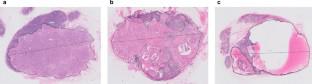
Digital pathology uses digitized images for cancer research. We aimed to assess morphometric parameters using digital pathology for predicting recurrence in patients with papillary thyroid carcinoma (PTC) and lateral cervical lymph node (LN) metastasis. We analyzed 316 PTC patients and assessed the longest diameter and largest area of metastatic focus in LNs using a whole slide imaging scanner. In digital pathology assessment, the longest diameters and largest areas of metastatic foci in LNs were positively correlated with traditional optically measured diameters (R = 0.928 and R2 = 0.727, p < 0.001 and p < 0.001, respectively). The optimal cutoff diameter was 8.0 mm in both traditional microscopic (p = 0.009) and digital pathology (p = 0.016) evaluations, with significant differences in progression-free survival (PFS) observed at this cutoff (p = 0.006 and p = 0.002, respectively). The predictive area’s cutoff was 35.6 mm2 (p = 0.005), which significantly affected PFS (p = 0.015). Using an 8.0-mm cutoff in traditional microscopic evaluation and a 35.6-mm2 cutoff in digital pathology showed comparable predictive results using the proportion of variation explained (PVE) methods (2.6% vs. 2.4%). Excluding cases with predominant cystic changes in LNs, the largest metastatic areas by digital pathology had the highest PVE at 3.9%. Furthermore, high volume of LN metastasis (p = 0.001), extranodal extension (p = 0.047), and high ratio of metastatic LNs (p = 0.006) were associated with poor prognosis. Both traditional microscopic and digital pathology evaluations effectively measured the longest diameter of metastatic foci in LNs. Moreover, digital pathology offers limited advantages in predicting PFS of patients with lateral cervical LN metastasis of PTC, especially those without predominant cystic changes in LNs.
中文翻译:
数字病理学对甲状腺乳头状癌颈侧淋巴结转移的形态学分析
数字病理学使用数字化图像进行癌症研究。我们的目的是使用数字病理学评估形态参数,以预测甲状腺乳头状癌(PTC)和颈侧淋巴结(LN)转移患者的复发。我们分析了 316 名 PTC 患者,并使用全玻片成像扫描仪评估了 LN 转移灶的最长直径和最大面积。在数字病理学评估中,LN 中转移灶的最长直径和最大面积与传统光学测量直径呈正相关(分别为R = 0.928 和R 2 = 0.727,p < 0.001 和p < 0.001)。传统显微镜 ( p = 0.009) 和数字病理学 ( p = 0.016) 评估中的最佳截止直径均为 8.0 mm,在此截止值下观察到的无进展生存期 (PFS) 存在显着差异(p = 0.006 和p = 0.002,分别)。预测区域的截止值为 35.6 mm 2 ( p = 0.005),这显着影响 PFS ( p = 0.015)。在传统显微评估中使用 8.0 毫米截止值,在数字病理学中使用 35.6 毫米2截止值,使用变异解释比例 (PVE) 方法显示出可比的预测结果(2.6% 与 2.4%)。排除淋巴结中主要囊性改变的病例,数字病理学显示的最大转移区域的 PVE 最高,为 3.9%。此外,大量的淋巴结转移(p = 0.001)、结外扩散(p = 0.047)和高的转移淋巴结比例(p = 0.006)与不良预后相关。传统的显微和数字病理学评估都有效测量了淋巴结转移灶的最长直径。此外,数字病理学在预测 PTC 颈侧侧淋巴结转移患者的 PFS 方面优势有限,尤其是那些淋巴结无明显囊性改变的患者。


 京公网安备 11010802027423号