Nature Protocols ( IF 14.8 ) Pub Date : 2023-12-12 , DOI: 10.1038/s41596-023-00912-w Benjamin W. Gregor , Mackenzie E. Coston , Ellen M. Adams , Joy Arakaki , Antoine Borensztejn , Thao P. Do , Margaret A. Fuqua , Amanda Haupt , Melissa C. Hendershott , Winnie Leung , Irina A. Mueller , Aditya Nath , Angelique M. Nelson , Susanne M. Rafelski , Emmanuel E. Sanchez , Madison J. Swain-Bowden , W. Joyce Tang , Derek J. Thirstrup , Winfried Wiegraebe , Brian P. Whitney , Calysta Yan , Ruwanthi N. Gunawardane , Nathalie Gaudreault
|
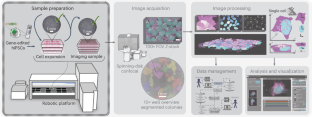
To produce abundant cell culture samples to generate large, standardized image datasets of human induced pluripotent stem (hiPS) cells, we developed an automated workflow on a Hamilton STAR liquid handler system. This was developed specifically for culturing hiPS cell lines expressing fluorescently tagged proteins, which we have used to study the principles by which cells establish and maintain robust dynamic localization of cellular structures. This protocol includes all details for the maintenance, passage and seeding of cells, as well as Matrigel coating of 6-well plastic plates and 96-well optical-grade, glass plates. We also developed an automated image-based hiPS cell colony segmentation and feature extraction pipeline to streamline the process of predicting cell count and selecting wells with consistent morphology for high-resolution three-dimensional (3D) microscopy. The imaging samples produced with this protocol have been used to study the integrated intracellular organization and cell-to-cell variability of hiPS cells to train and develop deep learning-based label-free predictions from transmitted-light microscopy images and to develop deep learning-based generative models of single-cell organization. This protocol requires some experience with robotic equipment. However, we provide details and source code to facilitate implementation by biologists less experienced with robotics. The protocol is completed in less than 10 h with minimal human interaction. Overall, automation of our cell culture procedures increased our imaging samples’ standardization, reproducibility, scalability and consistency. It also reduced the need for stringent culturist training and eliminated culturist-to-culturist variability, both of which were previous pain points of our original manual pipeline workflow.
中文翻译:
用于 3D 活细胞显微镜的自动化人类诱导多能干细胞培养和样品制备
为了产生丰富的细胞培养样本以生成人类诱导多能干 (hiPS) 细胞的大型标准化图像数据集,我们在 Hamilton STAR 液体处理系统上开发了自动化工作流程。这是专门为培养表达荧光标记蛋白的 hiPS 细胞系而开发的,我们用它来研究细胞建立和维持细胞结构的稳健动态定位的原理。该协议包括细胞维护、传代和接种的所有细节,以及 6 孔塑料板和 96 孔光学级玻璃板的基质胶涂层。我们还开发了一种基于图像的自动化 hiPS 细胞集落分割和特征提取流程,以简化预测细胞计数和选择具有一致形态的孔以进行高分辨率三维 (3D) 显微镜的过程。使用该协议生成的成像样本已用于研究 hiPS 细胞的集成细胞内组织和细胞间变异性,以训练和开发基于透射光显微镜图像的基于深度学习的无标签预测,并开发深度学习-基于单细胞组织的生成模型。该协议需要一些机器人设备的经验。但是,我们提供详细信息和源代码,以方便机器人技术经验不足的生物学家实施。该协议在不到 10 小时内完成,并且人机交互最少。总体而言,我们的细胞培养程序的自动化提高了成像样品的标准化、可重复性、可扩展性和一致性。它还减少了对严格的培养者培训的需求,并消除了培养者之间的差异,这两者都是我们原来的手动管道工作流程之前的痛点。


 京公网安备 11010802027423号