Journal of Cardiovascular Translational Research ( IF 3.4 ) Pub Date : 2023-12-13 , DOI: 10.1007/s12265-023-10472-9 Yijin Wu , Wenying Peng , Siyao Chen , Xiaodong Zeng , Jiade Zhu , Ping Zhu
|
Abstract
Extracellular vesicles (EVs) derived from mouse bone marrow mesenchymal stem cells (mBMSCs) convey the CAV1 protein, influencing the TGF-β1/SMAD2/c-JUN pathway and thus the molecular mechanisms underlying myocardial fibrosis (MF) post-myocardial infarction (MI). Through various experimental methods, including transmission electron microscopy, Nanosight analysis, Western blot, ELISA, and qRT-PCR, we isolated, purified, and identified EVs originating from mBMSCs. Bioinformatics and experimental findings show a reduced expression of CAV1 in myocardial fibrosis tissue. Furthermore, our findings suggest that mBMSC-EVs can deliver CAV1 to cardiac fibroblasts (CFs) and that silencing CAV1 in mBMSC-EVs promotes CF fibrosis. In vivo studies further corroborated these findings. In conclusion, mBMSC-EVs mitigate myocardial fibrosis in MI mice by delivering the CAV1 protein, inhibiting the TGF-β1/SMAD2/c-JUN pathway.
Graphical Abstract
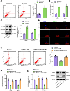
Molecular mechanism of mBMSC-EVs-CAV1-mediated TGF-β1/SMAD2/c-JUN axis in inhibiting cardiac fibroblast differentiation to improve MF after MI. mBMSC-EVs deliver CAV1 protein to CFs where the protein expression of CAV1 is upregulated upon hypoxia conditions. The TGF-β1/SMAD2 signaling pathway downstream of CAV1 is consequently inactivated, the transcription of c-JUN is inhibited, and transcription of SMAD2/c-JUN transcription complex target genes α-SMA and Collagen I is reduced. By this mechanism, CF fibrosis and apoptosis are suppressed in vitro and MF is ameliorated in MI mice.
中文翻译:
小鼠 BMSC 来源的细胞外囊泡中封装的 CAV1 蛋白通过阻断 TGF-β1/SMAD2/c-JUN 轴减轻心肌梗死后的心肌纤维化
摘要
源自小鼠骨髓间充质干细胞 (mBMSC) 的细胞外囊泡 (EV) 传递 CAV1 蛋白,影响 TGF-β1/SMAD2/c-JUN 通路,从而影响心肌梗死 (MI) 后心肌纤维化 (MF) 的分子机制)。通过各种实验方法,包括透射电子显微镜、Nanosight 分析、Western blot、ELISA 和 qRT-PCR,我们分离、纯化和鉴定了源自 mBMSC 的 EV。生物信息学和实验结果显示心肌纤维化组织中CAV1的表达降低。此外,我们的研究结果表明,mBMSC-EV 可以将 CAV1 传递给心脏成纤维细胞 (CF),并且沉默 mBMSC-EV 中的 CAV1 会促进 CF 纤维化。体内研究进一步证实了这些发现。总之,mBMSC-EVs 通过传递 CAV1 蛋白、抑制 TGF-β1/SMAD2/c-JUN 通路来减轻 MI 小鼠的心肌纤维化。
图形概要
mBMSC-EVs-CAV1介导的TGF-β1/SMAD2/c-JUN轴抑制心肌成纤维细胞分化改善MI后MF的分子机制。mBMSC-EV 将 CAV1 蛋白递送至 CF,其中 CAV1 的蛋白表达在缺氧条件下上调。CAV1下游的TGF-β1/SMAD2信号通路因此失活,c-JUN的转录受到抑制,SMAD2/c-JUN转录复合物靶基因α-SMA和胶原蛋白I的转录减少。通过这种机制,CF 纤维化和细胞凋亡在体外受到抑制,并且 MI 小鼠的 MF 得到改善。


 京公网安备 11010802027423号