当前位置:
X-MOL 学术
›
Eur. J. Neurosci.
›
论文详情
Our official English website, www.x-mol.net, welcomes your feedback! (Note: you will need to create a separate account there.)
Analysis of potassium ion diffusion from neurons to capillaries: Effects of astrocyte endfeet geometry
European Journal of Neuroscience ( IF 3.698 ) Pub Date : 2023-12-20 , DOI: 10.1111/ejn.16232 Sara Djurich 1 , Timothy W. Secomb 1
European Journal of Neuroscience ( IF 3.698 ) Pub Date : 2023-12-20 , DOI: 10.1111/ejn.16232 Sara Djurich 1 , Timothy W. Secomb 1
Affiliation
|
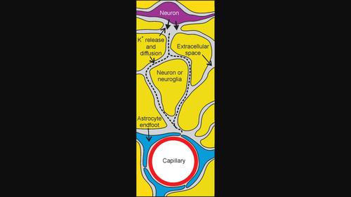
Neurovascular coupling (NVC) refers to a local increase in cerebral blood flow in response to increased neuronal activity. Mechanisms of communication between neurons and blood vessels remain unclear. Astrocyte endfeet almost completely cover cerebral capillaries, suggesting that astrocytes play a role in NVC by releasing vasoactive substances near capillaries. An alternative hypothesis is that direct diffusion through the extracellular space of potassium ions (K+) released by neurons contributes to NVC. Here, the goal is to determine whether astrocyte endfeet present a barrier to K+ diffusion from neurons to capillaries. Two simplified 2D geometries of extracellular space, clefts between endfeet, and perivascular space are used: (i) a source 1 μm from a capillary; (ii) a neuron 15 μm from a capillary. K+ release is modelled as a step increase in [K+] at the outer boundary of the extracellular space. The time-dependent diffusion equation is solved numerically. In the first geometry, perivascular [K+] approaches its final value within 0.05 s. Decreasing endfeet cleft width or increasing perivascular space width slows the rise in [K+]. In the second geometry, the increase in perivascular [K+] occurs within 0.5 s and is insensitive to changes in cleft width or perivascular space width. Predicted levels of perivascular [K+] are sufficient to cause vasodilation, and the rise time is within the time for flow increase in NVC. These results suggest that direct diffusion of K+ through the extracellular space is a possible NVC signalling mechanism.
中文翻译:

钾离子从神经元到毛细血管的扩散分析:星形胶质细胞端足几何形状的影响
神经血管耦合(NVC)是指因神经元活动增加而导致的脑血流量局部增加。神经元和血管之间的通讯机制仍不清楚。星形胶质细胞的端足几乎完全覆盖了脑毛细血管,这表明星形胶质细胞通过在毛细血管附近释放血管活性物质而在NVC中发挥作用。另一种假设是,神经元释放的钾离子 (K + )通过细胞外空间直接扩散有助于 NVC。此处的目标是确定星形胶质细胞终足是否对 K +从神经元到毛细血管的扩散存在障碍。使用两种简化的细胞外空间、端脚之间的裂缝和血管周围空间的二维几何形状:(i)距离毛细管 1 μm 的源; (ii) 距离毛细血管 15 μm 的神经元。 K +释放被建模为细胞外空间外边界处[K + ]的逐步增加。数值求解随时间变化的扩散方程。在第一个几何结构中,血管周围 [K + ] 在 0.05 秒内接近其最终值。减小足末裂宽度或增加血管周围间隙宽度会减慢 [K + ]的上升。在第二种几何结构中,血管周围 [K + ]的增加发生在 0.5 秒内,并且对裂隙宽度或血管周围空间宽度的变化不敏感。血管周围[K + ]的预测水平足以引起血管舒张,并且上升时间在NVC中流量增加的时间内。这些结果表明 K +通过细胞外空间的直接扩散是一种可能的 NVC 信号传导机制。
更新日期:2023-12-20
中文翻译:
钾离子从神经元到毛细血管的扩散分析:星形胶质细胞端足几何形状的影响
神经血管耦合(NVC)是指因神经元活动增加而导致的脑血流量局部增加。神经元和血管之间的通讯机制仍不清楚。星形胶质细胞的端足几乎完全覆盖了脑毛细血管,这表明星形胶质细胞通过在毛细血管附近释放血管活性物质而在NVC中发挥作用。另一种假设是,神经元释放的钾离子 (K + )通过细胞外空间直接扩散有助于 NVC。此处的目标是确定星形胶质细胞终足是否对 K +从神经元到毛细血管的扩散存在障碍。使用两种简化的细胞外空间、端脚之间的裂缝和血管周围空间的二维几何形状:(i)距离毛细管 1 μm 的源; (ii) 距离毛细血管 15 μm 的神经元。 K +释放被建模为细胞外空间外边界处[K + ]的逐步增加。数值求解随时间变化的扩散方程。在第一个几何结构中,血管周围 [K + ] 在 0.05 秒内接近其最终值。减小足末裂宽度或增加血管周围间隙宽度会减慢 [K + ]的上升。在第二种几何结构中,血管周围 [K + ]的增加发生在 0.5 秒内,并且对裂隙宽度或血管周围空间宽度的变化不敏感。血管周围[K + ]的预测水平足以引起血管舒张,并且上升时间在NVC中流量增加的时间内。这些结果表明 K +通过细胞外空间的直接扩散是一种可能的 NVC 信号传导机制。


 京公网安备 11010802027423号