当前位置:
X-MOL 学术
›
Eur. J. Nerosci.
›
论文详情
Our official English website, www.x-mol.net, welcomes your feedback! (Note: you will need to create a separate account there.)
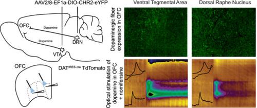
Characterization of dopaminergic projections from the ventral tegmental area and the dorsal raphe nucleus to the orbital frontal cortex
European Journal of Neroscience ( IF 3.4 ) Pub Date : 2023-12-28 , DOI: 10.1111/ejn.16230 Duncan J. Noble 1 , Aida Mohammadkhani 1 , Min Qiao 1 , Stephanie L. Borgland 1
European Journal of Neroscience ( IF 3.4 ) Pub Date : 2023-12-28 , DOI: 10.1111/ejn.16230 Duncan J. Noble 1 , Aida Mohammadkhani 1 , Min Qiao 1 , Stephanie L. Borgland 1
Affiliation
|
The orbitofrontal cortex (OFC) is a key node in the cortico-limbic-striatal circuitry that influences decision-making guided by the relative value of outcomes. Midbrain dopamine from either the ventral tegmental area (VTA) or the dorsal raphe nucleus (DRN) has the potential to modulate OFC neurons; however, it is unknown at what concentrations these terminals release dopamine. Male and female adult dopamine transporter (DAT)IRES-Cre–tdTomato mice were injected with AAV2/8-EF1a-DIO-eYFP into either the DRN or the VTA or the retrograde label cholera toxin B (CTB) 488 in the medial or lateral OFC. We quantified co-expression of CTB 488 or enhanced yellow fluorescent protein (eYFP) with tdTomato fluorescence in VTA or DRN and eYFP fibre density in the medial or lateral OFC. Both VTA and DRN dopamine neurons project to either the medial OFC or the lateral OFC, with greater expression of fibres in the medial OFC. Using fast-scan cyclic voltammetry, we detected optogenetically evoked dopamine from channelrhodopsin 2 (ChR2)-expressing VTA or DRN dopamine terminals in either the medial OFC or the lateral OFC. We assessed if optical stimulation of dopamine from the VTA or the DRN onto the medial OFC could alter layer V pyramidal neuronal firing; however, we did not observe a change in firing at stimulation parameters that evoked dopamine release from either projection even though bath application of dopamine with the monoamine transporter inhibitor, nomifensine, decreased firing. In summary, dopaminergic neurons from the VTA or the DRN project to the OFC and release submicromolar dopamine in the medial and lateral OFC.
中文翻译:

从腹侧被盖区和中缝背核到眶额皮质的多巴胺能投射的表征
眶额皮质(OFC)是皮质-边缘-纹状体回路中的关键节点,它影响由结果的相对价值引导的决策。来自腹侧被盖区 (VTA) 或中缝背核 (DRN) 的中脑多巴胺具有调节 OFC 神经元的潜力;然而,尚不清楚这些末端释放多巴胺的浓度。将 AAV2/8-EF1a-DIO-eYFP 注射到雄性和雌性成年多巴胺转运蛋白 (DAT) IRES-Cre –tdTomato 小鼠的 DRN 或 VTA 中,或在内侧或外侧注射逆行标签霍乱毒素 B (CTB) 488 OFC。我们量化了 CTB 488 或增强型黄色荧光蛋白 (eYFP) 与 tdTomato 荧光在 VTA 或 DRN 中的共表达以及内侧或外侧 OFC 中的 eYFP 纤维密度。 VTA 和 DRN 多巴胺神经元均投射到内侧 OFC 或外侧 OFC,其中内侧 OFC 中的纤维表达较多。使用快速扫描循环伏安法,我们检测到内侧 OFC 或外侧 OFC 中表达通道视紫红质 2 (ChR2) 的 VTA 或 DRN 多巴胺末端的光遗传学诱发的多巴胺。我们评估了从 VTA 或 DRN 到内侧 OFC 的多巴胺光刺激是否可以改变 V 层锥体神经元放电;然而,我们没有观察到刺激参数的放电变化,这些参数会引起任一投射的多巴胺释放,即使使用单胺转运蛋白抑制剂诺米芬辛沐浴应用多巴胺会减少放电。总之,来自 VTA 或 DRN 的多巴胺能神经元投射到 OFC,并在内侧和外侧 OFC 释放亚微摩尔多巴胺。
更新日期:2023-12-28
中文翻译:
从腹侧被盖区和中缝背核到眶额皮质的多巴胺能投射的表征
眶额皮质(OFC)是皮质-边缘-纹状体回路中的关键节点,它影响由结果的相对价值引导的决策。来自腹侧被盖区 (VTA) 或中缝背核 (DRN) 的中脑多巴胺具有调节 OFC 神经元的潜力;然而,尚不清楚这些末端释放多巴胺的浓度。将 AAV2/8-EF1a-DIO-eYFP 注射到雄性和雌性成年多巴胺转运蛋白 (DAT) IRES-Cre –tdTomato 小鼠的 DRN 或 VTA 中,或在内侧或外侧注射逆行标签霍乱毒素 B (CTB) 488 OFC。我们量化了 CTB 488 或增强型黄色荧光蛋白 (eYFP) 与 tdTomato 荧光在 VTA 或 DRN 中的共表达以及内侧或外侧 OFC 中的 eYFP 纤维密度。 VTA 和 DRN 多巴胺神经元均投射到内侧 OFC 或外侧 OFC,其中内侧 OFC 中的纤维表达较多。使用快速扫描循环伏安法,我们检测到内侧 OFC 或外侧 OFC 中表达通道视紫红质 2 (ChR2) 的 VTA 或 DRN 多巴胺末端的光遗传学诱发的多巴胺。我们评估了从 VTA 或 DRN 到内侧 OFC 的多巴胺光刺激是否可以改变 V 层锥体神经元放电;然而,我们没有观察到刺激参数的放电变化,这些参数会引起任一投射的多巴胺释放,即使使用单胺转运蛋白抑制剂诺米芬辛沐浴应用多巴胺会减少放电。总之,来自 VTA 或 DRN 的多巴胺能神经元投射到 OFC,并在内侧和外侧 OFC 释放亚微摩尔多巴胺。


 京公网安备 11010802027423号