SN Comprehensive Clinical Medicine Pub Date : 2024-01-10 , DOI: 10.1007/s42399-023-01637-3 Marco Pace , Marco Moretti , Simone Maria Tierno , Alessandro Dario Mazzotta , Andrea Felice Ferroni , Marco Di Giovan Paolo , Valeria Gianfreda , Salvatore Bianca , Apostolos Barbarosos , Carlo Eugenio Vitelli , Michelangelo Boninfante , Francesco Stipa
|
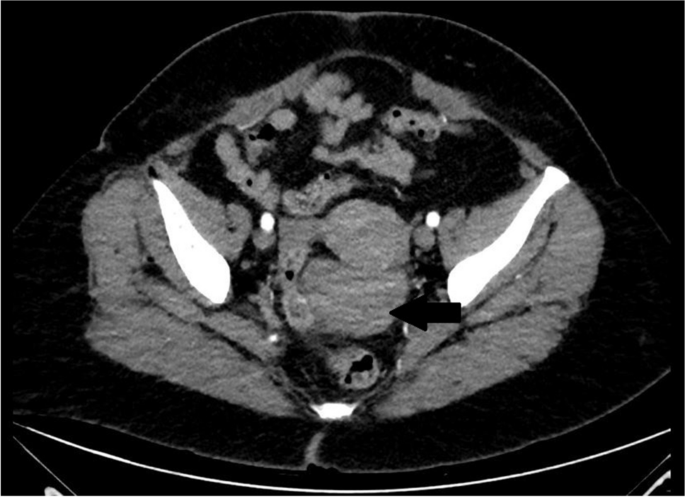
Leiomyomas are benign tumors, mostly located in the uterus. The pelvic localization is quite rare, and it is associated with unusual growth patterns. It is important to make an adequate differential diagnosis between malignant and benign retroperitoneal neoplasm because treatment is different. When it is not possible to have a precise preoperative diagnosis, a laparoscopic or laparotomy surgical tumorectomy is often required. To obtain a certain diagnosis, the goal of surgery is ensuring the complete excision of neoplasms and preservation of urination, defecation, and sexual function. We report a rare case of a 58-year-old woman who underwent a laparoscopic tumorectomy for a pelvic retroperitoneal leiomyoma. The patient reported occasional episodes of dull pain in the pelvic region. Pelvic contrast CT scan and magnetic resonance imaging (MRI) showed a retroperitoneal solid mass in contiguity with the posterior wall of the uterine body-isthmus, to be referred to as a pedunculated uterine fibroma strictly posteriorly adherent to the sigma. She first underwent to explorative laparoscopy by a gynecologist who did not find any uterine mass. The patient was subsequently admitted to the department of general surgery and has done a second operative laparoscopy which highlighted the presence of an extra-peritoneal para-rectal mass which was completely excised. The histological examination of tumor indicated that it was a leiomyoma. The postoperative course was uneventful, and the patient was discharged in III post-operative day (POD).
中文翻译:
腹腔镜肿瘤切除术治疗罕见盆腔腹膜后平滑肌瘤:病例报告
平滑肌瘤是良性肿瘤,大多数位于子宫。骨盆定位相当罕见,并且与不寻常的生长模式有关。由于治疗方法不同,对恶性和良性腹膜后肿瘤进行充分的鉴别诊断非常重要。当无法进行精确的术前诊断时,通常需要进行腹腔镜或剖腹手术肿瘤切除术。为了获得一定的诊断,手术的目标是确保肿瘤的完全切除并保留排尿、排便和性功能。我们报道了一名 58 岁女性的罕见病例,她因盆腔腹膜后平滑肌瘤接受了腹腔镜肿瘤切除术。患者报告骨盆区域偶尔出现钝痛。盆腔对比CT扫描和磁共振成像(MRI)显示腹膜后实性肿块与子宫体峡部后壁相邻,被称为带蒂子宫纤维瘤,严格向后粘附于西格玛。她首先接受了妇科医生的腹腔镜检查,没有发现任何子宫肿块。该患者随后被送往普通外科,并进行了第二次手术腹腔镜检查,结果显示存在腹膜外直肠旁肿块,该肿块已被完全切除。肿瘤的组织学检查表明其为平滑肌瘤。术后过程很顺利,患者在术后第三天(POD)出院。


 京公网安备 11010802027423号