Acta Neuropathologica ( IF 12.7 ) Pub Date : 2024-01-24 , DOI: 10.1007/s00401-023-02682-x Swenja Gödicke , Catena Kresbach , Max Ehlert , Denise Obrecht , Lea Altendorf , Karoline Hack , Katja von Hoff , Helena Carén , Viktoria Melcher , Kornelius Kerl , Bernhard Englinger , Mariella Filbin , Kristian W. Pajtler , Johannes Gojo , Torsten Pietsch , Stefan Rutkowski , Ulrich Schüller
|
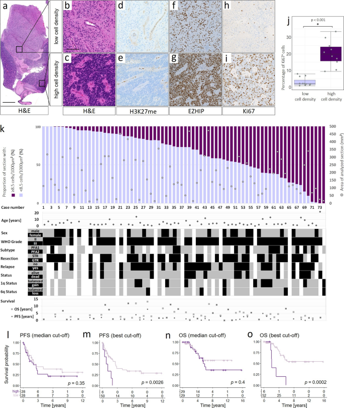
Posterior fossa type A (PF-EPN-A, PFA) ependymoma are aggressive tumors that mainly affect children and have a poor prognosis. Histopathology shows significant intratumoral heterogeneity, ranging from loose tissue to often sharply demarcated, extremely cell-dense tumor areas. To determine molecular differences in morphologically different areas and to understand their clinical significance, we analyzed 113 PF-EPN-A samples, including 40 corresponding relapse samples. Cell-dense areas ranged from 0 to 100% of the tumor area and displayed a higher proportion of proliferating tumor cells (p < 0.01). Clinically, cell density was associated with poor progression-free and overall survival (pPFS = 0.0026, pOS < 0.01). Molecularly, tumor areas with low and high cell density showed diverging DNA methylation profiles regarding their similarity to distinct previously discovered PF-EPN-A subtypes in 9/21 cases. Prognostically relevant chromosomal changes at 1q and 6q showed spatial heterogeneity within single tumors and were significantly enriched in cell-dense tumor areas as shown by single-cell RNA (scRNA)-sequencing as well as copy number profiling and fluorescence in situ hybridization (FISH) analyses of different tumor areas. Finally, spatial transcriptomics revealed cell-dense areas of different tumors to be more similar than various different areas of the same tumor. High-density areas distinctly overexpressed genes encoding histone proteins, WNT5A, TGFB1, or IGF2. Relapsing tumors displayed a higher proportion of cell-dense areas (p = 0.036), a change in PF-EPN-A methylation subtypes (13/32 patients), and novel chromosome 1q gains and 6q losses (12/32 cases) compared to corresponding primary tumors. Our data suggest that PF-EPN-A ependymomas habor a previously unrecognized intratumoral heterogeneity with clinical implications, which has to be accounted for when selecting diagnostic material, inter alia, by histological evaluation of the proportion of cell-dense areas.
中文翻译:
PFA 室管膜瘤的临床相关分子特征表现出瘤内异质性并与肿瘤形态相关
A 型后颅窝(PF-EPN-A、PFA)室管膜瘤是一种侵袭性肿瘤,主要影响儿童,预后不良。组织病理学显示显着的肿瘤内异质性,范围从疏松的组织到通常界限分明、细胞极其密集的肿瘤区域。为了确定形态不同区域的分子差异并了解其临床意义,我们分析了 113 个 PF-EPN-A 样本,其中包括 40 个相应的复发样本。细胞密集区域占肿瘤面积的 0% 至 100%,并且显示出较高比例的增殖肿瘤细胞 ( p < 0.01)。临床上,细胞密度与较差的无进展生存期和总生存期相关(p PFS = 0.0026,p OS < 0.01)。从分子角度来看,低细胞密度和高细胞密度的肿瘤区域与之前在 9/21 例中发现的不同 PF-EPN-A 亚型的相似性表现出不同的 DNA 甲基化谱。单细胞 RNA (scRNA) 测序以及拷贝数分析和荧光原位杂交 (FISH) 显示,1q 和 6q 处与预后相关的染色体变化显示出单个肿瘤内的空间异质性,并且在细胞密集的肿瘤区域中显着富集不同肿瘤区域的分析。最后,空间转录组学揭示了不同肿瘤的细胞密集区域比同一肿瘤的各个不同区域更相似。高密度区域明显过度表达编码组蛋白、WNT5A、TGFB1 或 IGF2 的基因。 与之前相比,复发肿瘤显示出更高比例的细胞密集区域(p = 0.036)、PF-EPN-A 甲基化亚型的变化(13/32 例患者)以及新的染色体 1q 增加和 6q 丢失(12/32 例)相应的原发肿瘤。我们的数据表明,PF-EPN-A 室管膜瘤具有以前未被认识到的具有临床意义的瘤内异质性,在选择诊断材料时必须考虑到这一点,尤其是通过细胞密集区域比例的组织学评估。


 京公网安备 11010802027423号