当前位置:
X-MOL 学术
›
Cytoskeleton
›
论文详情
Our official English website, www.x-mol.net, welcomes your feedback! (Note: you will need to create a separate account there.)
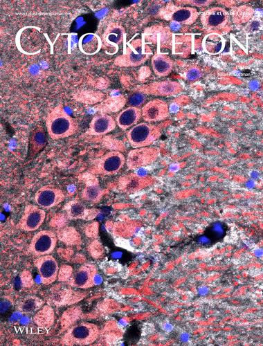
Inner Back Cover Image
Cytoskeleton ( IF 2.9 ) Pub Date : 2024-01-22 , DOI: 10.1002/cm.21831
Cytoskeleton ( IF 2.9 ) Pub Date : 2024-01-22 , DOI: 10.1002/cm.21831
|
ON THE INNER BACK COVER: Distribution of tau in the adult rat hippocampus (CA3 region shown). The tissue section was dephosphorylated and then stained with Tau1 antibody (white), MAP2 (red) and DAPI (blue). Tau is normally found at abundant levels within the soma, dendrites, axons and some nuclei of neurons (pink indicates colocalization between Tau1 and MAP2 signal).
中文翻译:

内封底图片
内封底:成年大鼠海马体中 tau 蛋白的分布(显示 CA3 区域)。将组织切片去磷酸化,然后用 Tau1 抗体(白色)、MAP2(红色)和 DAPI(蓝色)染色。Tau 通常在神经元的体细胞、树突、轴突和一些细胞核内含量丰富(粉色表示 Tau1 和 MAP2 信号共定位)。
更新日期:2024-01-25
中文翻译:
内封底图片
内封底:成年大鼠海马体中 tau 蛋白的分布(显示 CA3 区域)。将组织切片去磷酸化,然后用 Tau1 抗体(白色)、MAP2(红色)和 DAPI(蓝色)染色。Tau 通常在神经元的体细胞、树突、轴突和一些细胞核内含量丰富(粉色表示 Tau1 和 MAP2 信号共定位)。


 京公网安备 11010802027423号