当前位置:
X-MOL 学术
›
Acta Pharm. Sin. B
›
论文详情
Our official English website, www.x-mol.net, welcomes your feedback! (Note: you will need to create a separate account there.)
Enhanced optical imaging and fluorescent labeling for visualizing drug molecules within living organisms
Acta Pharmaceutica Sinica B ( IF 14.5 ) Pub Date : 2024-02-05 , DOI: 10.1016/j.apsb.2024.01.018 Ting Sun , Huanxin Zhao , Luyao Hu , Xintian Shao , Zhiyuan Lu , Yuli Wang , Peixue Ling , Yubo Li , Kewu Zeng , Qixin Chen
Acta Pharmaceutica Sinica B ( IF 14.5 ) Pub Date : 2024-02-05 , DOI: 10.1016/j.apsb.2024.01.018 Ting Sun , Huanxin Zhao , Luyao Hu , Xintian Shao , Zhiyuan Lu , Yuli Wang , Peixue Ling , Yubo Li , Kewu Zeng , Qixin Chen
|
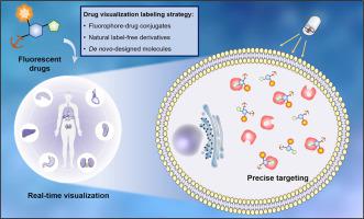
The visualization of drugs in living systems has become key techniques in modern therapeutics. Recent advancements in optical imaging technologies and molecular design strategies have revolutionized drug visualization. At the subcellular level, super-resolution microscopy has allowed exploration of the molecular landscape within individual cells and the cellular response to drugs. Moving beyond subcellular imaging, researchers have integrated multiple modes, like optical near-infrared II imaging, to study the complex spatiotemporal interactions between drugs and their surroundings. By combining these visualization approaches, researchers gain supplementary information on physiological parameters, metabolic activity, and tissue composition, leading to a comprehensive understanding of drug behavior. This review focuses on cutting-edge technologies in drug visualization, particularly fluorescence imaging, and the main types of fluorescent molecules used. Additionally, we discuss current challenges and prospects in targeted drug research, emphasizing the importance of multidisciplinary cooperation in advancing drug visualization. With the integration of advanced imaging technology and molecular design, drug visualization has the potential to redefine our understanding of pharmacology, enabling the analysis of drug micro-dynamics in subcellular environments from new perspectives and deepening pharmacological research to the levels of the cell and organelles.
中文翻译:

增强光学成像和荧光标记,用于可视化生物体内的药物分子
生命系统中药物的可视化已成为现代治疗学的关键技术。光学成像技术和分子设计策略的最新进展彻底改变了药物可视化。在亚细胞水平上,超分辨率显微镜可以探索单个细胞内的分子景观以及细胞对药物的反应。除了亚细胞成像之外,研究人员还集成了光学近红外 II 成像等多种模式来研究药物与其周围环境之间复杂的时空相互作用。通过结合这些可视化方法,研究人员可以获得有关生理参数、代谢活动和组织组成的补充信息,从而全面了解药物行为。本综述重点关注药物可视化的前沿技术,特别是荧光成像,以及所使用的荧光分子的主要类型。此外,我们讨论了靶向药物研究当前的挑战和前景,强调多学科合作在推进药物可视化方面的重要性。随着先进成像技术与分子设计的结合,药物可视化有可能重新定义我们对药理学的理解,使我们能够从新的角度分析亚细胞环境中的药物微动力学,将药理学研究深化到细胞和细胞器水平。
更新日期:2024-02-05
中文翻译:
增强光学成像和荧光标记,用于可视化生物体内的药物分子
生命系统中药物的可视化已成为现代治疗学的关键技术。光学成像技术和分子设计策略的最新进展彻底改变了药物可视化。在亚细胞水平上,超分辨率显微镜可以探索单个细胞内的分子景观以及细胞对药物的反应。除了亚细胞成像之外,研究人员还集成了光学近红外 II 成像等多种模式来研究药物与其周围环境之间复杂的时空相互作用。通过结合这些可视化方法,研究人员可以获得有关生理参数、代谢活动和组织组成的补充信息,从而全面了解药物行为。本综述重点关注药物可视化的前沿技术,特别是荧光成像,以及所使用的荧光分子的主要类型。此外,我们讨论了靶向药物研究当前的挑战和前景,强调多学科合作在推进药物可视化方面的重要性。随着先进成像技术与分子设计的结合,药物可视化有可能重新定义我们对药理学的理解,使我们能够从新的角度分析亚细胞环境中的药物微动力学,将药理学研究深化到细胞和细胞器水平。


 京公网安备 11010802027423号