Neurotoxicity Research ( IF 3.7 ) Pub Date : 2024-02-13 , DOI: 10.1007/s12640-024-00687-2 Xue Cheng , Zhetan Ren , Huiyang Jia , Gang Wang
|
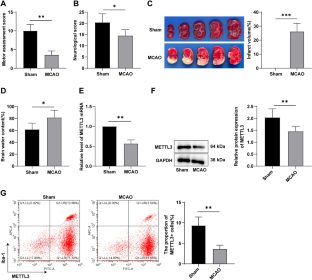
Cerebral ischemic stroke (CIS) is the main cause of disability. METTL3 is implicated in CIS, and we explored its specific mechanism. Middle cerebral artery occlusion (MCAO) rat model and oxygen–glucose deprivation/reperfusion (OGD/R) HAPI cell model were established and treated with LV-METTL3 or DAA, oe-METTL3, miR-335-3p mimics, or DAA, to assess their effects on MCAO rat neurological and motor function, cerebral infarction area, brain water content, microglial activation, blood–brain barrier (BBB) permeability, and NLRP3 inflammasome activation. METTL3, pri-miR-335-3p, mature miR-335-3p, and miR-335-3p mRNA levels were assessed by RT-qPCR; M1/M2 microglial phenotype proportion and M1/M2 microglia ratio, inflammatory factor levels, and m6A modification were assessed. MCAO rats manifested cerebral ischemia injury. METTL3 was under-expressed in CIS. METTL3 overexpression inhibited microglial activation and M1 polarization and BBB permeability in MCAO rats and inhibited OGD/R-induced microglial activation and reduced M1 polarization. METTL3 regulated miR-335-3p expression and inhibited NLRP3 inflammasome activation. m6A methylation inhibition averted METTL3’s effects on NLRP3 activation, thus promoting microglial activation in OGD/R-induced cells and METTL3’s effects on BBB permeability in MCAO rats. Briefly, METTL3 regulated miR-335-3p expression through RNA m6A methylation and inhibited NLRP3 inflammasome activation, thus repressing microglial activation, BBB permeability, and protecting against CIS.
中文翻译:
METTL3 通过 m6A 甲基化修饰调节 NLRP3 炎症小体介导脑缺血性中风中的小胶质细胞活化和血脑屏障通透性
脑缺血性中风(CIS)是导致残疾的主要原因。 METTL3与CIS有关,我们探讨了其具体机制。建立大脑中动脉闭塞(MCAO)大鼠模型和氧糖剥夺/再灌注(OGD/R)HAPI细胞模型,并用LV-METTL3或DAA、oe-METTL3、miR-335-3p模拟物或DAA处理,以评估它们对 MCAO 大鼠神经和运动功能、脑梗塞面积、脑含水量、小胶质细胞活化、血脑屏障 (BBB) 通透性和 NLRP3 炎性体活化的影响。通过 RT-qPCR 评估 METTL3、pri-miR-335-3p、成熟 miR-335-3p 和 miR-335-3p mRNA 水平;评估了 M1/M2 小胶质细胞表型比例和 M1/M2 小胶质细胞比率、炎症因子水平和 m6A 修饰。 MCAO大鼠表现为脑缺血损伤。 METTL3 在 CIS 中表达不足。 METTL3 过表达抑制 MCAO 大鼠中的小胶质细胞活化、M1 极化和 BBB 通透性,并抑制 OGD/R 诱导的小胶质细胞活化并减少 M1 极化。 METTL3 调节 miR-335-3p 表达并抑制 NLRP3 炎性体激活。 m6A 甲基化抑制避免了 METTL3 对 NLRP3 激活的影响,从而促进 OGD/R 诱导细胞中的小胶质细胞激活以及 METTL3 对 MCAO 大鼠中 BBB 通透性的影响。简而言之,METTL3 通过 RNA m6A 甲基化调节 miR-335-3p 表达并抑制 NLRP3 炎性体激活,从而抑制小胶质细胞激活、BBB 通透性并防止 CIS。


 京公网安备 11010802027423号