Nature Cell Biology ( IF 21.3 ) Pub Date : 2024-02-20 , DOI: 10.1038/s41556-024-01358-2 Tobias Roider , Marc A. Baertsch , Donnacha Fitzgerald , Harald Vöhringer , Berit J. Brinkmann , Felix Czernilofsky , Mareike Knoll , Laura Llaó-Cid , Anna Mathioudaki , Bianca Faßbender , Maxime Herbon , Tobias Lautwein , Peter-Martin Bruch , Nora Liebers , Christian M. Schürch , Verena Passerini , Marc Seifert , Alexander Brobeil , Gunhild Mechtersheimer , Carsten Müller-Tidow , Oliver Weigert , Martina Seiffert , Garry P. Nolan , Wolfgang Huber , Sascha Dietrich
|
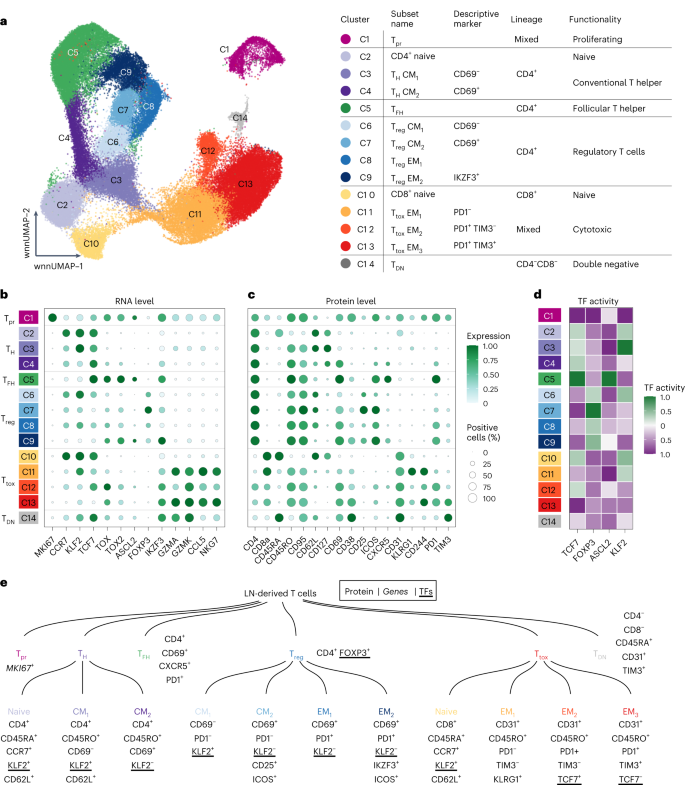
The redirection of T cells has emerged as an attractive therapeutic principle in B cell non-Hodgkin lymphoma (B-NHL). However, a detailed characterization of lymphoma-infiltrating T cells across B-NHL entities is missing. Here we present an in-depth T cell reference map of nodal B-NHL, based on cellular indexing of transcriptomes and epitopes, T cell receptor sequencing, flow cytometry and multiplexed immunofluorescence applied to 101 lymph nodes from patients with diffuse large B cell, mantle cell, follicular or marginal zone lymphoma, and from healthy controls. This multimodal resource revealed quantitative and spatial aberrations of the T cell microenvironment across and within B-NHL entities. Quantitative differences in PD1+ TCF7− cytotoxic T cells, T follicular helper cells or IKZF3+ regulatory T cells were linked to their clonal expansion. The abundance of PD1+ TCF7− cytotoxic T cells was associated with poor survival. Our study portrays lymphoma-infiltrating T cells with unprecedented comprehensiveness and provides a unique resource for the investigation of lymphoma biology and prognosis.
中文翻译:
多模式和空间解析分析可识别结节 B 细胞淋巴瘤实体中 T 细胞浸润的不同模式
T 细胞的重定向已成为 B 细胞非霍奇金淋巴瘤 (B-NHL) 的一个有吸引力的治疗原则。然而,B-NHL 实体中淋巴瘤浸润 T 细胞的详细特征却缺失。在这里,我们展示了淋巴结 B-NHL 的深入 T 细胞参考图,基于转录组和表位的细胞索引、T 细胞受体测序、流式细胞术和多重免疫荧光,应用于弥漫性大 B 细胞、套膜患者的 101 个淋巴结细胞、滤泡或边缘区淋巴瘤,以及来自健康对照。这种多模式资源揭示了 B-NHL 实体之间和内部 T 细胞微环境的数量和空间畸变。PD1 + TCF7 -细胞毒性 T 细胞、滤泡辅助 T 细胞或 IKZF3 +调节性 T 细胞的数量差异与其克隆扩张有关。PD1 + TCF7 -细胞毒性 T 细胞的丰度与生存率低相关。我们的研究以前所未有的全面性描绘了淋巴瘤浸润性 T 细胞,并为淋巴瘤生物学和预后的研究提供了独特的资源。


 京公网安备 11010802027423号