Our official English website, www.x-mol.net, welcomes your feedback! (Note: you will need to create a separate account there.)
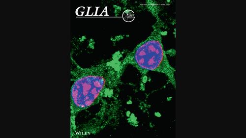
Cover Image, Volume 72, Issue 4
Glia ( IF 6.2 ) Pub Date : 2024-02-15 , DOI: 10.1002/glia.24399 Ioanna Zota , Konstantina Chanoumidou , Ioannis Charalampopoulos , Achille Gravanis
Glia ( IF 6.2 ) Pub Date : 2024-02-15 , DOI: 10.1002/glia.24399 Ioanna Zota , Konstantina Chanoumidou , Ioannis Charalampopoulos , Achille Gravanis
|
Cover Illustration: Confocal microscopy image of in vitro cultured proliferative oligodendrocyte progenitor cells (OPCs) under neurodegenerative conditions induced by the presence of toxic Amyloid-beta 1-42. The OPCs were immunostained for cellspecific PDGFRa marker (in green) and for Ki67 marker (in red) indicating proliferation. Hoechst dye (in blue) was used to stain nuclei of the total number of cells. (See Zota, I, et al, https://doi.org/10.1002/glia.24505)
中文翻译:

封面图片,第 72 卷,第 4 期
封面插图:在有毒的淀粉样蛋白-β 1-42 的存在诱导的神经退行性条件下,体外培养的增殖性少突胶质细胞祖细胞 (OPC) 的共聚焦显微镜图像。OPC 对细胞特异性 PDGFRa 标记(绿色)和 Ki67 标记(红色)进行免疫染色,表明增殖。Hoechst 染料(蓝色)用于对细胞总数的细胞核进行染色。(参见 Zota, I 等人,https://doi.org/10.1002/glia.24505)
更新日期:2024-02-20
中文翻译:
封面图片,第 72 卷,第 4 期
封面插图:在有毒的淀粉样蛋白-β 1-42 的存在诱导的神经退行性条件下,体外培养的增殖性少突胶质细胞祖细胞 (OPC) 的共聚焦显微镜图像。OPC 对细胞特异性 PDGFRa 标记(绿色)和 Ki67 标记(红色)进行免疫染色,表明增殖。Hoechst 染料(蓝色)用于对细胞总数的细胞核进行染色。(参见 Zota, I 等人,https://doi.org/10.1002/glia.24505)


 京公网安备 11010802027423号