Doklady Biochemistry and Biophysics ( IF 0.8 ) Pub Date : 2024-02-20 , DOI: 10.1134/s1607672923700709 Y. V. Khramtsov , A. V. Ulasov , T. N. Lupanova , G. P. Georgiev , A. S. Sobolev
|
Abstract
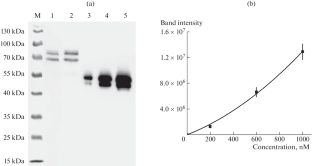
Two eukaryotic cell lines, A549 and A431, with stable expression of the nucleocapsid protein (N-protein) of the SARS-CoV-2 virus fused with the red fluorescent protein mRuby3 were obtained. Using microscopy, the volumes of the cytoplasm and nucleus were determined for these cells. Using quantitative immunoblotting techniques, the concentrations of the N-mRuby3 fusion protein in their cytoplasm were assessed. They were 19 and 9 μM for A549 and A431 cells, respectively. Using these concentrations, the initial rate of N-protein degradation in the studied cells was estimated from the decrease in cell fluorescence. In A549 and A431 cells, it was the same (84 nM per hour). The approach of quantitatively describing the degradation process can be applied to analyze the effectiveness of a wide class of antiviral drugs that cause degradation of viral proteins.
中文翻译:
在新型模块化纳米转运蛋白的影响下,稳定表达 SARS-CoV-2 病毒的 N 蛋白在细胞中降解的定量描述
摘要
获得了两种真核细胞系 A549 和 A431,它们稳定表达与红色荧光蛋白 mRuby3 融合的 SARS-CoV-2 病毒核衣壳蛋白(N 蛋白)。使用显微镜测定这些细胞的细胞质和细胞核的体积。使用定量免疫印迹技术,评估了细胞质中 N-mRuby3 融合蛋白的浓度。A549 和 A431 细胞的浓度分别为 19 和 9 μM。使用这些浓度,根据细胞荧光的减少来估计所研究细胞中 N 蛋白降解的初始速率。在 A549 和 A431 细胞中,它是相同的(84 nM 每小时)。定量描述降解过程的方法可用于分析导致病毒蛋白降解的多种抗病毒药物的有效性。


 京公网安备 11010802027423号