Developmental Cell ( IF 11.8 ) Pub Date : 2024-02-14 , DOI: 10.1016/j.devcel.2024.01.025 Hua Tian , Presha Rajbhandari , Jay Tarolli , Aubrianna M. Decker , Taruna V. Neelakantan , Tina Angerer , Fereshteh Zandkarimi , Helen Remotti , Gilles Frache , Nicholas Winograd , Brent R. Stockwell
|
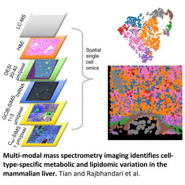
Spatial single-cell omics provides a readout of biochemical processes. It is challenging to capture the transient lipidome/metabolome from cells in a native tissue environment. We employed water gas cluster ion beam secondary ion mass spectrometry imaging ([H2O]n>28K-GCIB-SIMS) at ≤3 μm resolution using a cryogenic imaging workflow. This allowed multiple biomolecular imaging modes on the near-native-state liver at single-cell resolution. Our workflow utilizes desorption electrospray ionization (DESI) to build a reference map of metabolic heterogeneity and zonation across liver functional units at tissue level. Cryogenic dual-SIMS integrated metabolomics, lipidomics, and proteomics in the same liver lobules at single-cell level, characterizing the cellular landscape and metabolic states in different cell types. Lipids and metabolites classified liver metabolic zones, cell types and subtypes, highlighting the power of spatial multi-omics at high spatial resolution for understanding celluar and biomolecular organizations in the mammalian liver.
中文翻译:
多模态质谱成像识别哺乳动物肝脏中细胞类型特异性代谢和脂质组学变异
空间单细胞组学提供生化过程的读数。从天然组织环境中的细胞中捕获瞬时脂质组/代谢组具有挑战性。我们使用低温成像工作流程,采用水气团簇离子束二次离子质谱成像 ([H 2 O] n>2 8K -GCIB-SIMS),分辨率≤3 μm。这允许在接近天然状态的肝脏上以单细胞分辨率进行多种生物分子成像模式。我们的工作流程利用解吸电喷雾电离 (DESI) 来构建组织水平上肝功能单位代谢异质性和分区的参考图。低温双 SIMS 在单细胞水平上整合了同一肝小叶中的代谢组学、脂质组学和蛋白质组学,表征了不同细胞类型的细胞景观和代谢状态。脂质和代谢物对肝脏代谢区、细胞类型和亚型进行了分类,突出了高空间分辨率的空间多组学对于了解哺乳动物肝脏中的细胞和生物分子组织的能力。


 京公网安备 11010802027423号