当前位置:
X-MOL 学术
›
J. Mol. Cell. Cardiol.
›
论文详情
Our official English website, www.x-mol.net, welcomes your feedback! (Note: you will need to create a separate account there.)
Adipocyte-mediated electrophysiological remodeling of human stem cell - derived cardiomyocytes
Journal of Molecular and Cellular Cardiology ( IF 5 ) Pub Date : 2024-02-10 , DOI: 10.1016/j.yjmcc.2024.02.002 Justin Morrissette-McAlmon , William R. Xu , Roald Teuben , Kenneth R. Boheler , Leslie Tung
Journal of Molecular and Cellular Cardiology ( IF 5 ) Pub Date : 2024-02-10 , DOI: 10.1016/j.yjmcc.2024.02.002 Justin Morrissette-McAlmon , William R. Xu , Roald Teuben , Kenneth R. Boheler , Leslie Tung
|
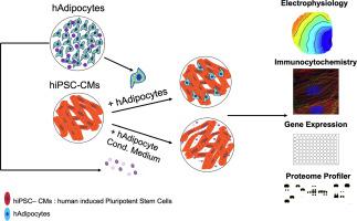
Adipocytes normally accumulate in the epicardial and pericardial layers around the human heart, but their infiltration into the myocardium can be proarrhythmic. Human adipose derived stem/stromal cells and human induced pluripotent stem cells (hiPSC) were differentiated, respectively into predominantly white fat-like adipocytes (hAdip) and ventricular cardiomyocytes (CMs). Adipocytes cultured in CM maintenance medium (CM medium) maintained their morphology, continued to express adipogenic markers, and retained clusters of intracellular lipid droplets. In contrast, hiPSC-CMs cultivated in adipogenic growth medium displayed abnormal cell morphologies and more clustering across the monolayer. Pre-plated hiPSC-CMs co-cultured in direct contact with hAdips in CM medium displayed prolonged action potential durations, increased triangulation, slowed conduction velocity, increased conduction velocity heterogeneity, and prolonged calcium transients. When hAdip-conditioned medium was added to monolayer cultures of hiPSC-CMs, results similar to those recorded with direct co-cultures were observed. Both co-culture and conditioned medium experiments resulted in increases in transcript abundance of SCN10A, CACNA1C, SLC8A1, and RYR2, with a decrease in KCNJ2. Human adipokine immunoblots revealed the presence of cytokines that were elevated in adipocyte-conditioned medium, including MCP-1, IL-6, IL-8 and CFD that could induce electrophysiological changes in cultured hiPSC-CMs. Co-culture of hiPSC-CMs with hAdips reveals a potentially pathogenic role of infiltrating human adipocytes on myocardial tissue. In the absence of structural changes, hAdip paracrine release alone is sufficient to cause CM electrophysiological dysfunction mirroring the co-culture conditions. These effects, mediated largely by paracrine mechanisms, could promote arrhythmias in the heart.
中文翻译:

脂肪细胞介导的人干细胞来源的心肌细胞的电生理重塑
脂肪细胞通常积聚在人类心脏周围的心外膜和心包层中,但它们渗入心肌可能会导致心律失常。人脂肪干细胞/基质细胞和人诱导多能干细胞(hiPSC)分别分化为主要为白色脂肪样脂肪细胞(hAdip)和心室心肌细胞(CM)。在CM维持培养基(CM培养基)中培养的脂肪细胞保持其形态,继续表达脂肪形成标记物,并保留细胞内脂滴簇。相比之下,在脂肪生长培养基中培养的 hiPSC-CM 显示出异常的细胞形态,并且在单层上聚集更多。在 CM 培养基中与 hAdips 直接接触共培养的预铺 hiPSC-CM 显示出延长的动作电位持续时间、增加的三角测量、减慢的传导速度、增加的传导速度异质性和延长的钙瞬变。当将 hAdip 条件培养基添加到 hiPSC-CM 的单层培养物中时,观察到与直接共培养物记录的结果相似的结果。共培养和条件培养基实验均导致 SCN10A、CACNA1C、SLC8A1 和 RYR2 转录本丰度增加,而 KCNJ2 转录本丰度减少。人类脂肪因子免疫印迹显示脂肪细胞条件培养基中存在升高的细胞因子,包括 MCP-1、IL-6、IL-8 和 CFD,它们可以诱导培养的 hiPSC-CM 中的电生理变化。 hiPSC-CM 与 hAdips 的共培养揭示了浸润人类脂肪细胞对心肌组织的潜在致病作用。在没有结构变化的情况下,单独 hAdip 旁分泌释放足以引起 CM 电生理功能障碍,反映了共培养条件。这些作用主要由旁分泌机制介导,可能会促进心律失常。
更新日期:2024-02-10
中文翻译:
脂肪细胞介导的人干细胞来源的心肌细胞的电生理重塑
脂肪细胞通常积聚在人类心脏周围的心外膜和心包层中,但它们渗入心肌可能会导致心律失常。人脂肪干细胞/基质细胞和人诱导多能干细胞(hiPSC)分别分化为主要为白色脂肪样脂肪细胞(hAdip)和心室心肌细胞(CM)。在CM维持培养基(CM培养基)中培养的脂肪细胞保持其形态,继续表达脂肪形成标记物,并保留细胞内脂滴簇。相比之下,在脂肪生长培养基中培养的 hiPSC-CM 显示出异常的细胞形态,并且在单层上聚集更多。在 CM 培养基中与 hAdips 直接接触共培养的预铺 hiPSC-CM 显示出延长的动作电位持续时间、增加的三角测量、减慢的传导速度、增加的传导速度异质性和延长的钙瞬变。当将 hAdip 条件培养基添加到 hiPSC-CM 的单层培养物中时,观察到与直接共培养物记录的结果相似的结果。共培养和条件培养基实验均导致 SCN10A、CACNA1C、SLC8A1 和 RYR2 转录本丰度增加,而 KCNJ2 转录本丰度减少。人类脂肪因子免疫印迹显示脂肪细胞条件培养基中存在升高的细胞因子,包括 MCP-1、IL-6、IL-8 和 CFD,它们可以诱导培养的 hiPSC-CM 中的电生理变化。 hiPSC-CM 与 hAdips 的共培养揭示了浸润人类脂肪细胞对心肌组织的潜在致病作用。在没有结构变化的情况下,单独 hAdip 旁分泌释放足以引起 CM 电生理功能障碍,反映了共培养条件。这些作用主要由旁分泌机制介导,可能会促进心律失常。


 京公网安备 11010802027423号