Cell and Tissue Research ( IF 3.6 ) Pub Date : 2024-02-22 , DOI: 10.1007/s00441-024-03879-6 Nick J. Spencer , Melinda A. Kyloh , Lee Travis , Timothy J. Hibberd
|
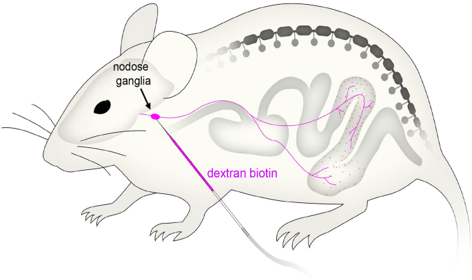
Understanding how the gut communicates with the brain, via sensory nerves, is of significant interest to medical science. Enteroendocrine cells (EEC) that line the mucosa of the gastrointestinal tract release neurochemicals, including the largest quantity of 5-hydroxytryptamine (5-HT). How the release of substances, like 5-HT, from enterochromaffin (EC) cells activates vagal afferent nerve endings is unresolved. We performed anterograde labelling from nodose ganglia in vivo and identified vagal afferent axons and nerve endings in the mucosa of whole-mount full-length preparations of mouse colon. We then determined the spatial relationship between mucosal-projecting vagal afferent nerve endings and EC cells in situ using 3D imaging. The mean distances between vagal afferent nerve endings in the mucosa, or nearest varicosities along vagal afferent axon branches, and the nearest EC cell were 29.6 ± 19.2 μm (n = 107, N = 6) and 25.7 ± 15.2 μm (n = 119, N = 6), respectively. No vagal afferent endings made close contacts with EC cells. The distances between EC cells and vagal afferent endings are many hundreds of times greater than known distances between pre- and post-synaptic membranes (typically 10–20 nm) that underlie synaptic transmission in vertebrates. The absence of any close physical contacts between 5-HT-containing EC cells and vagal afferent nerve endings in the mucosa leads to the inescapable conclusion that the mechanism by which 5-HT release from ECs in the colonic mucosa occurs in a paracrine fashion, to activate vagal afferents.
中文翻译:
小鼠结肠迷走神经传入神经末梢的识别及其与肠嗜铬细胞的空间关系
了解肠道如何通过感觉神经与大脑进行交流对于医学界具有重要意义。胃肠道粘膜上的肠内分泌细胞 (EEC) 释放神经化学物质,包括最大量的 5-羟色胺 (5-HT)。肠嗜铬细胞 (EC) 释放 5-HT 等物质如何激活迷走神经传入神经末梢尚未解决。我们对体内结状神经节进行了顺行标记,并在小鼠结肠的全长制备物的粘膜中鉴定了迷走神经传入轴突和神经末梢。然后,我们使用 3D 成像确定了粘膜投射迷走神经传入神经末梢和原位 EC 细胞之间的空间关系。粘膜迷走神经传入神经末梢或沿迷走神经传入轴突分支的最近静脉曲张与最近 EC 细胞之间的平均距离为 29.6 ± 19.2 μm ( n = 107,N = 6) 和 25.7 ± 15.2 μm ( n = 119,N = 6),分别。迷走神经传入末梢与 EC 细胞没有密切接触。EC 细胞和迷走神经传入末梢之间的距离比已知的脊椎动物突触传递基础的突触前膜和突触后膜之间的距离(通常为 10-20 nm)大数百倍。含 5-HT 的 EC 细胞与粘膜中的迷走神经传入神经末梢之间不存在任何紧密的物理接触,导致不可避免的结论,即结肠粘膜中的 EC 释放 5-HT 的机制以旁分泌方式发生,激活迷走神经传入。


 京公网安备 11010802027423号