当前位置:
X-MOL 学术
›
Brain Res.
›
论文详情
Our official English website, www.x-mol.net, welcomes your feedback! (Note: you will need to create a separate account there.)
Structural changes in spinal cord following optic neuritis: Insights from quantitative spinal MRI
Brain Research ( IF 2.9 ) Pub Date : 2024-02-24 , DOI: 10.1016/j.brainres.2024.148830 Jiyuan Wang , Jing Huang , Zheng Sun , Huiqing Dong , Kuncheng Li , Jie Lu
Brain Research ( IF 2.9 ) Pub Date : 2024-02-24 , DOI: 10.1016/j.brainres.2024.148830 Jiyuan Wang , Jing Huang , Zheng Sun , Huiqing Dong , Kuncheng Li , Jie Lu
|
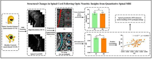
Previous studies have demonstrated that optic neuritis (ON) affects brain plasticity. However, whether ON affects the spinal cord remains unclear. We aimed to investigate the spinal cord changes in ON and their associations with disability. A total of 101 ON patients, and 41 healthy controls (HC) were retrospectively recruited. High-resolution imaging was conducted using a Magnetization Prepared Rapid Acquisition Gradient-Echo (MP-RAGE) sequence for T1-weighted images and an echo planar imaging (EPI) sequence for Diffusion Tensor Imaging (DTI) data collection. Additionally, patients' disability and cognitive impairment were evaluated using the Expanded Disability Status Scale (EDSS) and the Paced Auditory Serial Addition Test (PASAT), respectively. The quantitative spinal MRI was employed to examine the cross-sectional area (CSA) and diffusion indicators, with a specific focus on calculating the average values across the C2-C7 cervical spinal cord segments. CSA, fractional anisotropy (FA), mean diffusivity (MD), axial diffusivity (AD), and radial diffusivity (RD) were compared between groups. Correlation analyses were performed between CSA, diffusion indicators, and clinical variables. No significant differences were found in CSA between ON patients and HCs. MD (p = 0.007) and RD (p = 0.018) were increased in ON patients compared with HCs, and AD was decreased in ON (p = 0.013). The AD values of the ON patients were significantly positively correlated with PASAT scores (r = 0.37, p < 0.001). This study provided imaging evidence for DTI abnormalities in patients with ON. Spinal cord DTI can improve our knowledge of the path physiology of ON, and clinical progression.
中文翻译:

视神经炎后脊髓的结构变化:定量脊髓 MRI 的见解
先前的研究表明,视神经炎(ON)会影响大脑的可塑性。然而,ON是否影响脊髓仍不清楚。我们的目的是调查 ON 的脊髓变化及其与残疾的关系。回顾性招募了 101 名 ON 患者和 41 名健康对照 (HC)。使用用于 T1 加权图像的磁化准备快速采集梯度回波 (MP-RAGE) 序列和用于扩散张量成像 (DTI) 数据收集的回波平面成像 (EPI) 序列进行高分辨率成像。此外,还分别使用扩展残疾状态量表(EDSS)和节奏听觉连续附加测试(PASAT)评估患者的残疾和认知障碍。采用定量脊髓 MRI 检查横截面积 (CSA) 和弥散指标,特别关注计算 C2-C7 颈脊髓段的平均值。比较各组之间的 CSA、分数各向异性 (FA)、平均扩散率 (MD)、轴向扩散率 (AD) 和径向扩散率 (RD)。对 CSA、扩散指标和临床变量之间进行相关性分析。 ON 患者和 HC 之间的 CSA 没有发现显着差异。与 HC 相比,ON 患者的 MD (p = 0.007) 和 RD (p = 0.018) 增加,而 ON 患者的 AD 减少 (p = 0.013)。 ON患者的AD值与PASAT评分显着正相关(r = 0.37,p < 0.001)。这项研究为 ON 患者 DTI 异常提供了影像学证据。脊髓 DTI 可以提高我们对 ON 路径生理学和临床进展的了解。
更新日期:2024-02-24
中文翻译:
视神经炎后脊髓的结构变化:定量脊髓 MRI 的见解
先前的研究表明,视神经炎(ON)会影响大脑的可塑性。然而,ON是否影响脊髓仍不清楚。我们的目的是调查 ON 的脊髓变化及其与残疾的关系。回顾性招募了 101 名 ON 患者和 41 名健康对照 (HC)。使用用于 T1 加权图像的磁化准备快速采集梯度回波 (MP-RAGE) 序列和用于扩散张量成像 (DTI) 数据收集的回波平面成像 (EPI) 序列进行高分辨率成像。此外,还分别使用扩展残疾状态量表(EDSS)和节奏听觉连续附加测试(PASAT)评估患者的残疾和认知障碍。采用定量脊髓 MRI 检查横截面积 (CSA) 和弥散指标,特别关注计算 C2-C7 颈脊髓段的平均值。比较各组之间的 CSA、分数各向异性 (FA)、平均扩散率 (MD)、轴向扩散率 (AD) 和径向扩散率 (RD)。对 CSA、扩散指标和临床变量之间进行相关性分析。 ON 患者和 HC 之间的 CSA 没有发现显着差异。与 HC 相比,ON 患者的 MD (p = 0.007) 和 RD (p = 0.018) 增加,而 ON 患者的 AD 减少 (p = 0.013)。 ON患者的AD值与PASAT评分显着正相关(r = 0.37,p < 0.001)。这项研究为 ON 患者 DTI 异常提供了影像学证据。脊髓 DTI 可以提高我们对 ON 路径生理学和临床进展的了解。


 京公网安备 11010802027423号