Journal of Cardiovascular Translational Research ( IF 3.4 ) Pub Date : 2024-02-26 , DOI: 10.1007/s12265-024-10502-0 Belay Tesfamariam
|
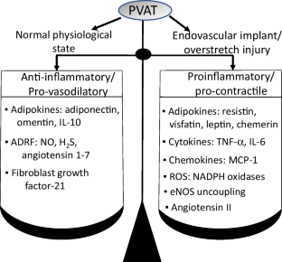
Following the placement of endovascular implants, perivascular adipose tissue (PVAT) becomes an early sensor of vascular injury to which it responds by undergoing phenotypic changes characterized by reduction in the secretion of adipocyte-derived relaxing factors and a shift to a proinflammatory and pro-contractile state. Thus, activated PVAT loses its anti-inflammatory function, secretes proinflammatory cytokines and chemokines, and generates reactive oxygen species, which are accompanied by differentiation of fibroblasts into myofibroblasts and proliferation of smooth muscle cells. These subsequently migrate into the intima, leading to intimal growth. In addition, periadventitial vasa vasorum undergoes neovascularization and functions as a portal for extravasation of inflammatory infiltrates and mobilization of PVAT resident stem/progenitor cells into the intima. This review focuses on the response of PVAT to endovascular intervention-induced injury and discusses potential therapeutic targets to suppress the PVAT-initiated pathways that mediate the formation of neointima.
Graphical Abstract
中文翻译:
血管内放置后血管周围脂肪组织对新内膜形成的影响
放置血管内植入物后,血管周围脂肪组织(PVAT)成为血管损伤的早期传感器,它通过经历表型变化来做出反应,其特征是脂肪细胞衍生的松弛因子的分泌减少以及向促炎和促收缩因子的转变状态。因此,活化的PVAT失去其抗炎功能,分泌促炎细胞因子和趋化因子,并产生活性氧,伴随着成纤维细胞向肌成纤维细胞的分化和平滑肌细胞的增殖。这些随后迁移到内膜中,导致内膜生长。此外,外膜周围的血管滋养管经历新血管形成,并作为炎症浸润外渗和动员PVAT驻留干细胞/祖细胞进入内膜的门户。本综述重点关注 PVAT 对血管内介入引起的损伤的反应,并讨论抑制 PVAT 启动介导新内膜形成途径的潜在治疗靶点。


 京公网安备 11010802027423号