Biomechanics and Modeling in Mechanobiology ( IF 3.5 ) Pub Date : 2024-02-28 , DOI: 10.1007/s10237-023-01794-3 Somaye Jafari , Joseph Park , Yongtao Lu , Joseph L. Demer
|
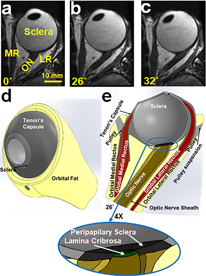
Details of the anatomy and behavior of the structures responsible for human eye movements have been extensively elaborated since the first modern biomechanical models were introduced. Based on these findings, a finite element model of human ocular adduction is developed based on connective anatomy and measured optic nerve (ON) properties, as well as active contractility of bilaminar extraocular muscles (EOMs), but incorporating the novel feature that globe translation is not otherwise constrained so that realistic kinematics can be simulated. Anatomy of the hemisymmetric model is defined by magnetic resonance imaging. The globe is modeled as suspended by anatomically realistic connective tissues, orbital fat, and contiguous ON. The model incorporates a material subroutine that implements active EOM contraction based on fiber twitch characteristics. Starting from the initial condition of 26° adduction, the medial rectus (MR) muscle was commanded to contract as the lateral rectus (LR) relaxed. We alternatively modeled absence or presence of orbital fat. During pursuit-like adduction from 26 to 32°, the globe translated 0.52 mm posteriorly and 0.1 mm medially with orbital fat present, but 1.2 mm posteriorly and 0.1 mm medially without fat. Maximum principal strains in the optic disk and peripapillary reached 0.05–0.06, and von-Mises stress 96 kPa. Tension in the MR orbital layer was ~ 24 g-force after 6° adduction, but only ~ 3 gm-f in the whole LR. This physiologically plausible simulation of EOM activation in an anatomically realistic globe suspensory system demonstrates that orbital connective tissues and fat are integral to the biomechanics of adduction, including loading by the ON.
中文翻译:
无约束球体平移的眼内收有限元模型
自从第一个现代生物力学模型被引入以来,负责人眼运动的结构的解剖学和行为细节已经得到了广泛的阐述。基于这些发现,基于结缔解剖学和测量的视神经 (ON) 特性以及双层眼外肌 (EOM) 的主动收缩性,开发了人眼内收的有限元模型,但结合了全球平移的新功能不受其他约束,以便可以模拟真实的运动学。半对称模型的解剖结构通过磁共振成像来定义。地球被建模为由解剖学上真实的结缔组织、眼眶脂肪和连续的 ON 悬浮而成。该模型包含一个材料子程序,可根据纤维抽搐特性实现主动 EOM 收缩。从26°内收的初始状态开始,当外直肌(LR)放松时,内直肌(MR)被命令收缩。我们另外模拟了眼眶脂肪的缺失或存在。在从 26° 到 32° 的追踪式内收过程中,眼球向后平移 0.52 毫米,向内平移 0.1 毫米,存在眼眶脂肪,但向后平移 1.2 毫米,向内平移 0.1 毫米,没有脂肪。视盘和视乳头周围的最大主应变达到0.05-0.06,von-Mises应力96 kPa。6°内收后,MR 轨道层的张力为约 24 g-f,但整个 LR 中的张力仅为约 3 gm-f。这种在解剖学上真实的球形悬吊系统中对 EOM 激活的生理学合理模拟表明,眼眶结缔组织和脂肪是内收生物力学不可或缺的一部分,包括 ON 的加载。


 京公网安备 11010802027423号