当前位置:
X-MOL 学术
›
Eur. J. Nerosci.
›
论文详情
Our official English website, www.x-mol.net, welcomes your feedback! (Note: you will need to create a separate account there.)
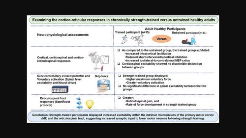
Strength‐trained adults demonstrate greater corticoreticular activation versus untrained controls
European Journal of Neroscience ( IF 3.4 ) Pub Date : 2024-02-29 , DOI: 10.1111/ejn.16297 Yonas Akalu 1, 2 , Jamie Tallent 1, 3 , Ashlyn K. Frazer 1 , Ummatul Siddique 1 , Mohamad Rostami 1 , Patrick Vallance 1 , Glyn Howatson 4, 5 , Simon Walker 6 , Dawson J. Kidgell 1
European Journal of Neroscience ( IF 3.4 ) Pub Date : 2024-02-29 , DOI: 10.1111/ejn.16297 Yonas Akalu 1, 2 , Jamie Tallent 1, 3 , Ashlyn K. Frazer 1 , Ummatul Siddique 1 , Mohamad Rostami 1 , Patrick Vallance 1 , Glyn Howatson 4, 5 , Simon Walker 6 , Dawson J. Kidgell 1
Affiliation
|
The rapid increase in strength following strength‐training involves neural adaptations, however, their specific localisation remains elusive. Prior focus on corticospinal responses prompts this study to explore the understudied cortical/subcortical adaptations, particularly cortico‐reticulospinal tract responses, comparing healthy strength‐trained adults to untrained peers. Fifteen chronically strength‐trained individuals (≥2 years of training, mean age: 24 ± 7 years) were compared with 11 age‐matched untrained participants (mean age: 26 ± 8 years). Assessments included maximal voluntary force (MVF), corticospinal excitability using transcranial magnetic stimulation (TMS), spinal excitability (cervicomedullary stimulation), voluntary activation (VA) and reticulospinal tract (RST) excitability, utilizing StartReact responses and ipsilateral motor‐evoked potentials (iMEPs) for the flexor carpi radialis muscle. Trained participants had higher normalized MVF (6.4 ± 1.1 N/kg) than the untrained participants (4.8 ± 1.3 N/kg) (p = .003). Intracortical facilitation was higher in the strength‐trained group (156 ± 49%) (p = .02), along with greater VA (98 ± 3.2%) (p = .002). The strength‐trained group displayed reduced short‐interval‐intracortical inhibition (88 ± 8.0%) compared with the untrained group (69 ± 17.5%) (p < .001). Strength‐trained individuals exhibited a greater normalized rate of force development (38.8 ± 10.1 N·s−1 /kg) (p < .009), greater reticulospinal gain (2.5 ± 1.4) (p = .02) and higher ipsilateral‐to‐contralateral MEP ratios compared with the untrained group (p = .03). Strength‐trained individuals displayed greater excitability within the intrinsic connections of the primary motor cortex and the RST. These results suggest greater synaptic input from the descending cortico‐reticulospinal tract to α‐motoneurons in strength‐trained individuals, thereby contributing to the observed increase in VA and MVF.
中文翻译:

与未经训练的对照组相比,经过力量训练的成年人表现出更强的皮质网状激活
力量训练后力量的快速增加涉及神经适应,然而,它们的具体定位仍然难以捉摸。先前对皮质脊髓反应的关注促使本研究探索尚未充分研究的皮质/皮质下适应性,特别是皮质网状脊髓束反应,将健康的接受过力量训练的成年人与未经训练的同龄人进行比较。将 15 名长期接受力量训练的个体(训练时间≥2 年,平均年龄:24 ± 7 岁)与 11 名年龄匹配的未训练参与者(平均年龄:26 ± 8 岁)进行比较。评估包括最大随意力 (MVF)、使用经颅磁刺激 (TMS) 的皮质脊髓兴奋性、脊髓兴奋性(颈髓刺激)、随意激活 (VA) 和网状脊髓束 (RST) 兴奋性,利用 StartReact 反应和同侧运动诱发电位 (iMEP) )对于桡侧腕屈肌。受过训练的参与者的标准化 MVF (6.4 ± 1.1 N/kg) 高于未经训练的参与者 (4.8 ± 1.3 N/kg)(p =.003)。力量训练组的皮质内促进较高(156 ± 49%)(p = .02),以及更大的 VA (98 ± 3.2%) (p = .002)。与未训练组 (69 ± 17.5%) 相比,力量训练组表现出短间隔皮质内抑制 (88 ± 8.0%) 减少(p < .001)。接受过力量训练的个体表现出更高的力量发展标准化速率(38.8 ± 10.1 N·s−1 /公斤) (p < .009), 更大的网状脊髓增益 (2.5 ± 1.4) (p = .02),与未训练组相比,同侧与对侧 MEP 比率更高(p = .03)。接受过力量训练的人在初级运动皮层和 RST 的内在联系中表现出更高的兴奋性。这些结果表明,在接受过力量训练的个体中,从皮质网状脊髓下降束到 α 运动神经元的突触输入更大,从而导致了观察到的 VA 和 MVF 的增加。
更新日期:2024-02-29
中文翻译:
与未经训练的对照组相比,经过力量训练的成年人表现出更强的皮质网状激活
力量训练后力量的快速增加涉及神经适应,然而,它们的具体定位仍然难以捉摸。先前对皮质脊髓反应的关注促使本研究探索尚未充分研究的皮质/皮质下适应性,特别是皮质网状脊髓束反应,将健康的接受过力量训练的成年人与未经训练的同龄人进行比较。将 15 名长期接受力量训练的个体(训练时间≥2 年,平均年龄:24 ± 7 岁)与 11 名年龄匹配的未训练参与者(平均年龄:26 ± 8 岁)进行比较。评估包括最大随意力 (MVF)、使用经颅磁刺激 (TMS) 的皮质脊髓兴奋性、脊髓兴奋性(颈髓刺激)、随意激活 (VA) 和网状脊髓束 (RST) 兴奋性,利用 StartReact 反应和同侧运动诱发电位 (iMEP) )对于桡侧腕屈肌。受过训练的参与者的标准化 MVF (6.4 ± 1.1 N/kg) 高于未经训练的参与者 (4.8 ± 1.3 N/kg)(


 京公网安备 11010802027423号