当前位置:
X-MOL 学术
›
Brain Res.
›
论文详情
Our official English website, www.x-mol.net, welcomes your feedback! (Note: you will need to create a separate account there.)
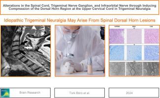
Alterations in the spinal cord, trigeminal nerve ganglion, and infraorbital nerve through inducing compression of the dorsal horn region at the upper cervical cord in trigeminal neuralgia
Brain Research ( IF 2.9 ) Pub Date : 2024-03-04 , DOI: 10.1016/j.brainres.2024.148842 Ülkü Türk Börü , Zülfikar Kadir Sarıtaş , Fatma Görücü Özbek , Cem Bölük , Hakan Acar , Yusuf Koç , Gökçe Zeytin Demiral
Brain Research ( IF 2.9 ) Pub Date : 2024-03-04 , DOI: 10.1016/j.brainres.2024.148842 Ülkü Türk Börü , Zülfikar Kadir Sarıtaş , Fatma Görücü Özbek , Cem Bölük , Hakan Acar , Yusuf Koç , Gökçe Zeytin Demiral
|
Idiopathic trigeminal neuralgia (TN) cases encountered frequently in daily practice indicate significant gaps that still need to be illuminated in the etiopathogenesis. In this study, a novel TN animal model was developed by compressing the dorsal horn (DH) of the upper cervical spinal cord. Eighteen rabbits were equally divided into three groups, namely control (CG), sham (SG), and spinal cord compression (SCC) groups. External pressure was applied to the left side at the C3 level in the SCC group. Dorsal hemilaminectomy was performed in the SG, and the operative side was closed without compression. No procedure was implemented in the control group. Samples from the SC, TG, and ION were taken after seven days. For the histochemical staining, damage and axons with myelin were scored using Hematoxylin and Eosin and Toluidine Blue, respectively. Immunohistochemistry, nuclei, apoptotic index, astrocyte activity, microglial labeling, and CD11b were evaluated. Mechanical allodynia was observed on the ipsilateral side in the SCC group. In addition, both the TG and ION were partially damaged from SC compression, which resulted in significant histopathological changes and increased the expression of all markers in both the SG and SCC groups compared to that in the CG. There was a notable increase in tissue damage, an increase in the number of apoptotic nuclei, an increase in the apoptotic index, an indication of astrocytic gliosis, and an upsurge in microglial cells. Significant increases were noted in the SG group, whereas more pronounced significant increases were observed in the SCC group. Transmission electron microscopy revealed myelin damage, mitochondrial disruption, and increased anchoring particles. Similar changes were observed to a lesser extent in the contralateral spinal cord. Ipsilateral trigeminal neuropathic pain was developed due to upper cervical SCC. The clinical finding is supported by immunohistochemical and ultrastructural changes. Thus, alterations in the DH due to compression of the upper cervical region should be considered as a potential cause of idiopathic TN.
中文翻译:

通过在三叉神经痛中诱导上颈髓背角区域受压来改变脊髓、三叉神经节和眶下神经
日常实践中经常遇到的特发性三叉神经痛 (TN) 病例表明,其发病机制仍存在重大空白,仍需阐明。在这项研究中,通过压缩上颈脊髓的背角(DH)开发了一种新型的 TN 动物模型。 18只兔子平均分为三组,即对照组(CG)、假手术组(SG)和脊髓压迫组(SCC)。对 SCC 组的左侧 C3 水平施加外部压力。在 SG 中进行背侧半椎板切除术,手术侧在不加压的情况下闭合。对照组未实施任何手术。 7 天后从 SC、TG 和 ION 采集样本。对于组织化学染色,分别使用苏木精、曙红和甲苯胺蓝对损伤和髓鞘质轴突进行评分。评估免疫组织化学、细胞核、凋亡指数、星形胶质细胞活性、小胶质细胞标记和 CD11b。 SCC 组同侧观察到机械性异常性疼痛。此外,TG和ION均因SC压缩而部分受损,导致SG和SCC组发生显着的组织病理学变化,并且与CG组相比,所有标记物的表达均增加。组织损伤显着增加,凋亡细胞核数量增加,凋亡指数增加,这是星形胶质细胞增生的迹象,以及小胶质细胞的激增。 SG 组中观察到显着增加,而 SCC 组中观察到更明显的显着增加。透射电子显微镜显示髓磷脂损伤、线粒体破坏和锚定颗粒增加。在对侧脊髓中也观察到较小程度的类似变化。同侧三叉神经性疼痛是由于上颈椎鳞状细胞癌引起的。免疫组织化学和超微结构变化支持了临床发现。因此,由于上颈部区域受压而导致的 DH 变化应被视为特发性 TN 的潜在原因。
更新日期:2024-03-04
中文翻译:
通过在三叉神经痛中诱导上颈髓背角区域受压来改变脊髓、三叉神经节和眶下神经
日常实践中经常遇到的特发性三叉神经痛 (TN) 病例表明,其发病机制仍存在重大空白,仍需阐明。在这项研究中,通过压缩上颈脊髓的背角(DH)开发了一种新型的 TN 动物模型。 18只兔子平均分为三组,即对照组(CG)、假手术组(SG)和脊髓压迫组(SCC)。对 SCC 组的左侧 C3 水平施加外部压力。在 SG 中进行背侧半椎板切除术,手术侧在不加压的情况下闭合。对照组未实施任何手术。 7 天后从 SC、TG 和 ION 采集样本。对于组织化学染色,分别使用苏木精、曙红和甲苯胺蓝对损伤和髓鞘质轴突进行评分。评估免疫组织化学、细胞核、凋亡指数、星形胶质细胞活性、小胶质细胞标记和 CD11b。 SCC 组同侧观察到机械性异常性疼痛。此外,TG和ION均因SC压缩而部分受损,导致SG和SCC组发生显着的组织病理学变化,并且与CG组相比,所有标记物的表达均增加。组织损伤显着增加,凋亡细胞核数量增加,凋亡指数增加,这是星形胶质细胞增生的迹象,以及小胶质细胞的激增。 SG 组中观察到显着增加,而 SCC 组中观察到更明显的显着增加。透射电子显微镜显示髓磷脂损伤、线粒体破坏和锚定颗粒增加。在对侧脊髓中也观察到较小程度的类似变化。同侧三叉神经性疼痛是由于上颈椎鳞状细胞癌引起的。免疫组织化学和超微结构变化支持了临床发现。因此,由于上颈部区域受压而导致的 DH 变化应被视为特发性 TN 的潜在原因。


 京公网安备 11010802027423号