当前位置:
X-MOL 学术
›
Brain Res.
›
论文详情
Our official English website, www.x-mol.net, welcomes your feedback! (Note: you will need to create a separate account there.)
Cerebral endothelial cells mediated enhancement of brain pericyte number and migration in oxygen-glucose deprivation involves the HIF-1α/PDGF-β signaling
Brain Research ( IF 2.9 ) Pub Date : 2024-03-05 , DOI: 10.1016/j.brainres.2024.148849 Shi-Na Song , Wen-Ping Dong , Xin-Xin Dong , Fang Guo , Lin Ren , Chang-Xin Li , Jian-Ming Wang
Brain Research ( IF 2.9 ) Pub Date : 2024-03-05 , DOI: 10.1016/j.brainres.2024.148849 Shi-Na Song , Wen-Ping Dong , Xin-Xin Dong , Fang Guo , Lin Ren , Chang-Xin Li , Jian-Ming Wang
|
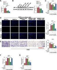
The present study focused on whether hypoxia-inducible factor-1alpha (HIF-1α) and platelet-derived factor-beta (PDGF-β) are involved in the crosstalk between brain microvascular endothelial cells (BMECs) and brain vascular pericytes (BVPs) under ischaemic-hypoxic conditions. Mono-cultures or co-cultures of BVPs and BMECs were made for the construction of the blood–brain barrier (BBB) model and then exposed to control and oxygen-glucose deprivation (OGD) conditions. BBB injury was determined by assessing the ability, apoptosis, and migration of BVPs and the transendothelial electrical resistance and horseradish peroxidase permeation of BMECs. Relative mRNA and protein levels of HIF-1α and PDGF-β, as well as tight junction proteins ZO-1 and claudin-5 were analyzed by western blotting, reverse transcription quantitative PCR, and/or immunofluorescence staining. Dual-luciferase reporter assays assessed the relationship between PDGF-β and HIF-1α. Co-culturing with BMECs alleviated OGD-induced reduction in BVP viability, elevation in BVP apoptosis, and repression in BVP migration. Co-culturing with BVPs protected against OGD-induced impairment on BMEC permeability. OGD-induced HIF-1α upregulation enhanced PDGF-β expression in mono-cultured BMECs and co-cultured BMECs with BVPs. Knockdown of HIF-1α impaired the effect of BMECs on BVPs under OGD conditions, and PDGFR-β silencing in BVPs blocked the crosstalk between BMECs and BVPs under OGD conditions. The crosstalk between BMECs and BVPs was implicated in OGD-induced BBB injury through the HIF-1α/PDGF-β signaling.
中文翻译:

氧葡萄糖剥夺中脑内皮细胞介导的脑周细胞数量和迁移的增强涉及 HIF-1α/PDGF-β 信号传导
本研究重点关注低氧诱导因子-1α(HIF-1α)和血小板衍生因子-β(PDGF-β)是否参与缺氧条件下脑微血管内皮细胞(BMEC)和脑血管周细胞(BVP)之间的串扰。缺血缺氧状况。 BVP 和 BMEC 的单一培养或共培养用于构建血脑屏障 (BBB) 模型,然后暴露于对照和氧糖剥夺 (OGD) 条件。通过评估 BVP 的能力、凋亡和迁移以及 BMEC 的跨内皮电阻和辣根过氧化物酶渗透来确定 BBB 损伤。通过蛋白质印迹、逆转录定量PCR和/或免疫荧光染色分析HIF-1α和PDGF-β以及紧密连接蛋白ZO-1和claudin-5的相对mRNA和蛋白水平。双荧光素酶报告基因测定评估了 PDGF-β 和 HIF-1α 之间的关系。与 BMEC 共培养减轻了 OGD 诱导的 BVP 活力降低、BVP 细胞凋亡升高和 BVP 迁移抑制。与 BVP 共培养可防止 OGD 引起的 BMEC 通透性损害。 OGD 诱导的 HIF-1α 上调增强了单培养 BMEC 以及与 BVP 共培养的 BMEC 中的 PDGF-β 表达。在 OGD 条件下,HIF-1α 的敲低削弱了 BMEC 对 BVP 的作用,而 BVP 中的 PDGFR-β 沉默则阻断了 OGD 条件下 BMEC 和 BVP 之间的串扰。 BMEC 和 BVP 之间的串扰通过 HIF-1α/PDGF-β 信号传导参与 OGD 诱导的 BBB 损伤。
更新日期:2024-03-05
中文翻译:
氧葡萄糖剥夺中脑内皮细胞介导的脑周细胞数量和迁移的增强涉及 HIF-1α/PDGF-β 信号传导
本研究重点关注低氧诱导因子-1α(HIF-1α)和血小板衍生因子-β(PDGF-β)是否参与缺氧条件下脑微血管内皮细胞(BMEC)和脑血管周细胞(BVP)之间的串扰。缺血缺氧状况。 BVP 和 BMEC 的单一培养或共培养用于构建血脑屏障 (BBB) 模型,然后暴露于对照和氧糖剥夺 (OGD) 条件。通过评估 BVP 的能力、凋亡和迁移以及 BMEC 的跨内皮电阻和辣根过氧化物酶渗透来确定 BBB 损伤。通过蛋白质印迹、逆转录定量PCR和/或免疫荧光染色分析HIF-1α和PDGF-β以及紧密连接蛋白ZO-1和claudin-5的相对mRNA和蛋白水平。双荧光素酶报告基因测定评估了 PDGF-β 和 HIF-1α 之间的关系。与 BMEC 共培养减轻了 OGD 诱导的 BVP 活力降低、BVP 细胞凋亡升高和 BVP 迁移抑制。与 BVP 共培养可防止 OGD 引起的 BMEC 通透性损害。 OGD 诱导的 HIF-1α 上调增强了单培养 BMEC 以及与 BVP 共培养的 BMEC 中的 PDGF-β 表达。在 OGD 条件下,HIF-1α 的敲低削弱了 BMEC 对 BVP 的作用,而 BVP 中的 PDGFR-β 沉默则阻断了 OGD 条件下 BMEC 和 BVP 之间的串扰。 BMEC 和 BVP 之间的串扰通过 HIF-1α/PDGF-β 信号传导参与 OGD 诱导的 BBB 损伤。


 京公网安备 11010802027423号