当前位置:
X-MOL 学术
›
Microsc. Res. Tech.
›
论文详情
Our official English website, www.x-mol.net, welcomes your feedback! (Note: you will need to create a separate account there.)
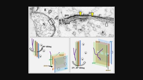
Electron‐translucency and partial defects of synaptic basal lamina in the electrocyte synapse of an electric ray (Narke japonica) in 3D embedment‐free section electron microscopy
Microscopy Research and Technique ( IF 2.5 ) Pub Date : 2024-03-10 , DOI: 10.1002/jemt.24534 Surang Chomphoo 1 , Hisatake Kondo 1, 2 , Wiphawi Hipkaeo 1
en‐face direction in embedment‐free section transmission electron microscopy (EFS‐TEM), and synaptic vesicles located close to the presynaptic membrane were seen through the synaptic basal lamina together with the presynaptic and postsynaptic membranes. This feature of translucency has the potential to analyze possible spatial interrelations in situ between bioactive molecules in the synaptic basal lamina and the synaptic vesicles in further studies. The synaptic basal lamina, appearing as an electron‐dense line sandwiched by two parallel lines representing the presynaptic and postsynaptic membranes in ultrathin sections cut right to the synaptic junctional plane in conventional TEM, was not fully continuous but randomly intermittent along its trajectory. Compatible with the intermittent line appearance, the en‐face 3D view in embedment‐free section TEM revealed for the first time partial irregular defects of the synaptic basal lamina. Considering the known functional significance of several molecules contained in the synaptic basal lamina in the maintenance and exertion of the synapse, its partial defects may not represent its rigid structural features, but its immature structure under remodeling or its dynamic changes in consistency such as the sol/gel transition, whose validity needs further examination.Research Highlights In embedment‐free section TEM, a 3D en‐face view of synaptic basal lamina in situ is reliably possible. The basal lamina en‐face is electron‐translucent, which makes it possible to analyze spatial interrelation between pre‐ and post‐synaptic components. Partial irregular defects in the basal lamina are revealed in Torpedo electrocytes, suggesting its remodeling or dynamic changes in consistency.
中文翻译:

3D 无嵌入切片电子显微镜中电鳐(Narke japonica)电细胞突触中突触基底层的电子半透明度和部分缺陷
研究发现,从电子角度观察时,电细胞的突触基底层在某种程度上是电子半透明的。面对 在无嵌入切片透射电子显微镜(EFS-TEM)中,通过突触基底层以及突触前膜和突触后膜可以看到靠近突触前膜的突触小泡。半透明的这一特征有可能在进一步的研究中分析突触基底层和突触小泡中生物活性分子之间可能的空间相互关系。在传统 TEM 中,突触基底层表现为一条电子密集线,夹在代表突触前和突触后膜的超薄切片中的两条平行线之间,该超薄切片直接切至突触连接平面,但突触基底层并不完全连续,而是沿其轨迹随机间歇。兼容断续线外观,面对 无嵌入切片 TEM 的 3D 视图首次揭示了突触基底层的部分不规则缺陷。考虑到突触基底层中包含的几种分子在维持和发挥突触方面已知的功能意义,其部分缺陷可能并不代表其刚性的结构特征,而是其重塑中的不成熟结构或其一致性的动态变化,例如溶胶/gel转变,其有效性有待进一步检验。 研究亮点 在免嵌入切片 TEM 中,可以可靠地获得突触基底层原位的 3D 正面视图。 基底层正面是电子半透明的,这使得分析突触前和突触后成分之间的空间相互关系成为可能。 鱼雷电细胞中发现基底层的部分不规则缺陷,表明其重塑或一致性的动态变化。
更新日期:2024-03-10
Microscopy Research and Technique ( IF 2.5 ) Pub Date : 2024-03-10 , DOI: 10.1002/jemt.24534 Surang Chomphoo 1 , Hisatake Kondo 1, 2 , Wiphawi Hipkaeo 1
Affiliation
|
中文翻译:
3D 无嵌入切片电子显微镜中电鳐(Narke japonica)电细胞突触中突触基底层的电子半透明度和部分缺陷


 京公网安备 11010802027423号