当前位置:
X-MOL 学术
›
Thorac. Cancer
›
论文详情
Our official English website, www.x-mol.net, welcomes your feedback! (Note: you will need to create a separate account there.)
Non‐small cell lung carcinoma with focal coexpression of thyroid transcription factor‐1 and Δ Np63/p40: A case report
Thoracic Cancer ( IF 2.9 ) Pub Date : 2024-03-13 , DOI: 10.1111/1759-7714.15271 Kazuhiro Terada 1 , Toshi Menju 2 , Horoshi Date 2 , Hironori Haga 1 , Akihiko Yoshizawa 1, 3
Thoracic Cancer ( IF 2.9 ) Pub Date : 2024-03-13 , DOI: 10.1111/1759-7714.15271 Kazuhiro Terada 1 , Toshi Menju 2 , Horoshi Date 2 , Hironori Haga 1 , Akihiko Yoshizawa 1, 3
Affiliation
|
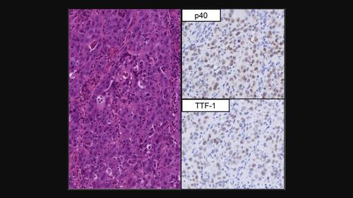
Most lung carcinomas are subtyped by their morphologies; however, immunohistochemistry is usually performed when it is difficult to determine. The most reliable antibodies for distinguishing lung adenocarcinoma from squamous cell carcinoma are thyroid transcription factor‐1 (TTF‐1) and p40 (ΔNp63). In general, these markers are mutually exclusive in their expression of lung primary carcinoma; however, a few cases of non‐small cell lung carcinoma (NSCLC) with coexpression of both markers have been reported. Examining a tissue microarray of 229 squamous cell carcinomas and 346 adenocarcinomas, we found one case of NSCLC with coexpression of TTF‐1 and p40. Herein, we present a 71‐year‐old man, who had a mass lesion in the left lung apex. A transbronchial lung biopsy was performed, revealing NSCLC. He underwent left upper segmentectomy and lymph node dissection. Macroscopically, the mass showed a white‐to‐tan solid tumor on the cut surface. Microscopically, the tumor was composed of polygonal tumor cells which had round and vesicular nuclei with prominent nucleoli. They had an abundant amount of cytoplasm, which was slightly eosinophilic or amphophilic. Multinucleated cells with atypical nuclear features were observed to be scattered in some areas. Multifocal necrosis and hemorrhage were also noted. Distinct squamous features and obvious glandular features were absent. Immunohistochemically, the most tumor cells were coexpressed positive for both TTF‐1 and p40. In our study, NSCLC with TTF‐1 and p40 coexpression is rare; therefore, it is necessary to obtain further data and examine similar cases to establish more precise definitions and clinicopathological features.
中文翻译:

甲状腺转录因子 1 和 Δ Np63/p40 局部共表达的非小细胞肺癌:一例报告
大多数肺癌根据其形态进行亚型分类。然而,当难以确定时,通常会进行免疫组化。区分肺腺癌和鳞状细胞癌最可靠的抗体是甲状腺转录因子-1 (TTF-1) 和 p40 (ΔNp63)。一般来说,这些标志物在肺原发性癌中的表达是相互排斥的;然而,已经报道了一些同时表达这两种标志物的非小细胞肺癌(NSCLC)病例。通过检查 229 个鳞状细胞癌和 346 个腺癌的组织微阵列,我们发现一例 NSCLC 共表达 TTF-1 和 p40。在此,我们介绍了一名 71 岁的男性,他的左肺尖有肿块病变。进行了经支气管肺活检,发现非小细胞肺癌。他接受了左上段切除术和淋巴结清扫术。肉眼可见,肿块切面呈白色至棕褐色的实体瘤。镜下观察,肿瘤由多角形肿瘤细胞组成,细胞核呈圆形、囊泡状,核仁突出。它们具有丰富的细胞质,呈轻微嗜酸性或两性。观察到核特征不典型的多核细胞分散在某些区域。还注意到多灶性坏死和出血。缺乏明显的鳞状特征和明显的腺体特征。免疫组织化学显示,大多数肿瘤细胞 TTF-1 和 p40 共表达阳性。在我们的研究中,TTF-1 和 p40 共表达的 NSCLC 很少见;因此,有必要获得进一步的数据并检查类似病例,以建立更精确的定义和临床病理特征。
更新日期:2024-03-13
中文翻译:
甲状腺转录因子 1 和 Δ Np63/p40 局部共表达的非小细胞肺癌:一例报告
大多数肺癌根据其形态进行亚型分类。然而,当难以确定时,通常会进行免疫组化。区分肺腺癌和鳞状细胞癌最可靠的抗体是甲状腺转录因子-1 (TTF-1) 和 p40 (ΔNp63)。一般来说,这些标志物在肺原发性癌中的表达是相互排斥的;然而,已经报道了一些同时表达这两种标志物的非小细胞肺癌(NSCLC)病例。通过检查 229 个鳞状细胞癌和 346 个腺癌的组织微阵列,我们发现一例 NSCLC 共表达 TTF-1 和 p40。在此,我们介绍了一名 71 岁的男性,他的左肺尖有肿块病变。进行了经支气管肺活检,发现非小细胞肺癌。他接受了左上段切除术和淋巴结清扫术。肉眼可见,肿块切面呈白色至棕褐色的实体瘤。镜下观察,肿瘤由多角形肿瘤细胞组成,细胞核呈圆形、囊泡状,核仁突出。它们具有丰富的细胞质,呈轻微嗜酸性或两性。观察到核特征不典型的多核细胞分散在某些区域。还注意到多灶性坏死和出血。缺乏明显的鳞状特征和明显的腺体特征。免疫组织化学显示,大多数肿瘤细胞 TTF-1 和 p40 共表达阳性。在我们的研究中,TTF-1 和 p40 共表达的 NSCLC 很少见;因此,有必要获得进一步的数据并检查类似病例,以建立更精确的定义和临床病理特征。


 京公网安备 11010802027423号