当前位置:
X-MOL 学术
›
ACS Biomater. Sci. Eng.
›
论文详情
Our official English website, www.x-mol.net, welcomes your feedback! (Note: you will need to create a separate account there.)
Formation Pattern Analysis of Spheroids Formed by a Droplet-Based Microfluidic System for Predicting the Aggressiveness of Tumor Cells
ACS Biomaterials Science & Engineering ( IF 5.8 ) Pub Date : 2024-03-14 , DOI: 10.1021/acsbiomaterials.4c00005 Sunghan Lee 1, 2 , Chang Jae Woo 2, 3 , Hyo-Il Jung 1, 4 , Ki Chang Nam 2 , Ji Seok Lim 5, 6 , Bong Seop Kwak 2, 6
ACS Biomaterials Science & Engineering ( IF 5.8 ) Pub Date : 2024-03-14 , DOI: 10.1021/acsbiomaterials.4c00005 Sunghan Lee 1, 2 , Chang Jae Woo 2, 3 , Hyo-Il Jung 1, 4 , Ki Chang Nam 2 , Ji Seok Lim 5, 6 , Bong Seop Kwak 2, 6
Affiliation
|
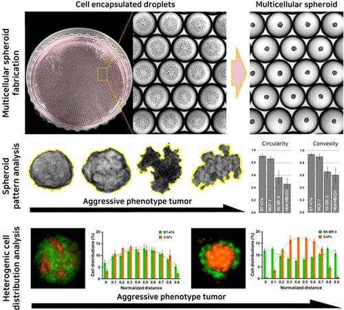
Examining tumor heterogeneity is essential for selecting an appropriate anticancer treatment for an individual. This study aimed to distinguish low- and high-aggressive tumor cells by analyzing the formation patterns of spheroids. The droplet-based microfluidic system was employed for the formation of each spheroid from four different subtypes of breast tumor cells. Additionally, heterotypic spheroids with T lymphocytes and cancer-associated fibroblasts (CAFs) were produced, and distinctions between low- and high-aggressive tumor cells were explored through the analysis of formation patterns using circularity, convexity, and cell distributions. In both homotypic spheroids and heterotypic spheroids with T lymphocytes, spheroids formed from low-aggressive tumor cells exhibited high circularity and convexity. On the other hand, spheroids formed from high-aggressive tumor cells had relatively low circularity and convexity. In the case of heterotypic spheroids with CAFs, circularity and convexity did not exhibit clear differences between low- and high-aggressive tumor cells, but distinct variations were observed in cell distributions. CAFs and low-aggressive tumor cells were evenly distributed, whereas the CAFs were predominantly located in the inner layer, and high-aggressive tumor cells were primarily located in the outer layer. This finding can offer valuable insights into predicting the aggressiveness of unknown tumor cells.
中文翻译:

基于液滴的微流体系统形成的球体的形成模式分析用于预测肿瘤细胞的侵袭性
检查肿瘤异质性对于为个体选择合适的抗癌治疗至关重要。本研究旨在通过分析球体的形成模式来区分低侵袭性和高侵袭性肿瘤细胞。基于液滴的微流体系统用于从四种不同亚型的乳腺肿瘤细胞形成每个球体。此外,还制备了含有 T 淋巴细胞和癌症相关成纤维细胞 (CAF) 的异型球体,并通过使用圆形、凸度和细胞分布分析形成模式来探索低侵袭性和高侵袭性肿瘤细胞之间的区别。在具有T淋巴细胞的同型球体和异型球体中,由低侵袭性肿瘤细胞形成的球体表现出高圆度和凸度。另一方面,由高侵袭性肿瘤细胞形成的球体具有相对较低的圆度和凸度。对于具有 CAF 的异型球体,低侵袭性和高侵袭性肿瘤细胞之间的圆形度和凸度没有表现出明显差异,但在细胞分布中观察到明显的变化。 CAF和低侵袭性肿瘤细胞分布均匀,而CAF主要位于内层,高侵袭性肿瘤细胞主要位于外层。这一发现可以为预测未知肿瘤细胞的侵袭性提供有价值的见解。
更新日期:2024-03-14
中文翻译:
基于液滴的微流体系统形成的球体的形成模式分析用于预测肿瘤细胞的侵袭性
检查肿瘤异质性对于为个体选择合适的抗癌治疗至关重要。本研究旨在通过分析球体的形成模式来区分低侵袭性和高侵袭性肿瘤细胞。基于液滴的微流体系统用于从四种不同亚型的乳腺肿瘤细胞形成每个球体。此外,还制备了含有 T 淋巴细胞和癌症相关成纤维细胞 (CAF) 的异型球体,并通过使用圆形、凸度和细胞分布分析形成模式来探索低侵袭性和高侵袭性肿瘤细胞之间的区别。在具有T淋巴细胞的同型球体和异型球体中,由低侵袭性肿瘤细胞形成的球体表现出高圆度和凸度。另一方面,由高侵袭性肿瘤细胞形成的球体具有相对较低的圆度和凸度。对于具有 CAF 的异型球体,低侵袭性和高侵袭性肿瘤细胞之间的圆形度和凸度没有表现出明显差异,但在细胞分布中观察到明显的变化。 CAF和低侵袭性肿瘤细胞分布均匀,而CAF主要位于内层,高侵袭性肿瘤细胞主要位于外层。这一发现可以为预测未知肿瘤细胞的侵袭性提供有价值的见解。


 京公网安备 11010802027423号