Nature ( IF 64.8 ) Pub Date : 2024-03-13 , DOI: 10.1038/s41586-024-07153-1 Makaía M. Papasergi-Scott , Guillermo Pérez-Hernández , Hossein Batebi , Yang Gao , Gözde Eskici , Alpay B. Seven , Ouliana Panova , Daniel Hilger , Marina Casiraghi , Feng He , Luis Maul , Peter Gmeiner , Brian K. Kobilka , Peter W. Hildebrand , Georgios Skiniotis
|
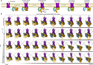
G-protein-coupled receptors (GPCRs) activate heterotrimeric G proteins by stimulating guanine nucleotide exchange in the Gα subunit1. To visualize this mechanism, we developed a time-resolved cryo-EM approach that examines the progression of ensembles of pre-steady-state intermediates of a GPCR–G-protein complex. By monitoring the transitions of the stimulatory Gs protein in complex with the β2-adrenergic receptor at short sequential time points after GTP addition, we identified the conformational trajectory underlying G-protein activation and functional dissociation from the receptor. Twenty structures generated from sequential overlapping particle subsets along this trajectory, compared to control structures, provide a high-resolution description of the order of main events driving G-protein activation in response to GTP binding. Structural changes propagate from the nucleotide-binding pocket and extend through the GTPase domain, enacting alterations to Gα switch regions and the α5 helix that weaken the G-protein–receptor interface. Molecular dynamics simulations with late structures in the cryo-EM trajectory support that enhanced ordering of GTP on closure of the α-helical domain against the nucleotide-bound Ras-homology domain correlates with α5 helix destabilization and eventual dissociation of the G protein from the GPCR. These findings also highlight the potential of time-resolved cryo-EM as a tool for mechanistic dissection of GPCR signalling events.
中文翻译:
GPCR 激活 G 蛋白的时间分辨冷冻电镜
G 蛋白偶联受体 (GPCR) 通过刺激 Gα 亚基中的鸟嘌呤核苷酸交换来激活异三聚体 G 蛋白1。为了可视化这一机制,我们开发了一种时间分辨冷冻电镜方法,用于检查 GPCR-G 蛋白复合物的前稳态中间体整体的进展。通过在添加 GTP 后的短连续时间点监测刺激性 G s蛋白与 β 2 -肾上腺素能受体复合物的转变,我们确定了 G 蛋白激活和与受体功能解离的构象轨迹。与对照结构相比,沿该轨迹由顺序重叠的粒子子集生成的 20 个结构提供了响应 GTP 结合而驱动 G 蛋白激活的主要事件顺序的高分辨率描述。结构变化从核苷酸结合口袋传播并延伸到 GTP 酶结构域,对 Gα 开关区域和 α5 螺旋进行改变,从而削弱 G 蛋白-受体界面。冷冻电镜轨迹中后期结构的分子动力学模拟支持,α-螺旋结构域相对于核苷酸结合的 Ras 同源结构域闭合时 GTP 的增强排序与 α5 螺旋不稳定以及 G 蛋白最终从 GPCR 解离相关。 。这些发现还强调了时间分辨冷冻电镜作为 GPCR 信号事件机械剖析工具的潜力。


 京公网安备 11010802027423号