Biomechanics and Modeling in Mechanobiology ( IF 3.5 ) Pub Date : 2024-03-15 , DOI: 10.1007/s10237-024-01835-5 Yasutaka Tobe , Takanobu Yagi , Koichi Kawamura , Kenta Suto , Yoichi Sawada , Yoshifumi Hayashi , Hirotaka Yoshida , Kazutoshi Nishitani , Yoshifumi Okada , Shigemi Kitahara , Mitsuo Umezu
|
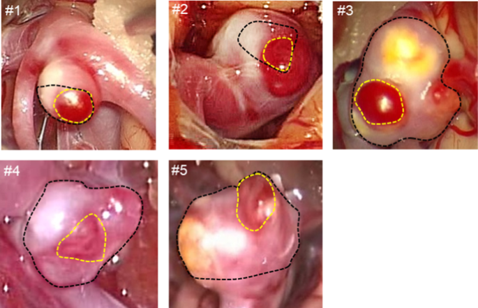
Aneurysmal rupture is associated with wall thinning, but the mechanism is poorly understood. This study aimed to characterize the three-dimensional wall-thickness distributions of unruptured intracranial aneurysms. Five aneurysmal tissues were investigated using micro-computed tomography. First, the wall thickness was related to the aneurysmal wall appearances during surgery. The median wall thicknesses of the translucent and non-translucent walls were 50.56 and 155.93 µm, respectively (p < 0.05) with significant variation in the non-translucent wall thicknesses (p < 0.05). The three-dimensional observations characterized the spatial variation of wall thicknesses. Thin walls showed a uniform thickness profile ranging from 10 to 40 µm, whereas thick walls presented a peaked thickness profile ranging from 300 to 500 µm. In transition walls, the profile undulated due to the formation of focal thin/thick spots. Overall, the aneurysmal wall thicknesses were strongly site-dependent and spatially varied by 10 to 40 times within individual cases. Aneurysmal walls are exposed to wall stress driven by blood pressure. In theory, the magnitude of wall stress is inversely proportional to wall thickness. Thus, the observed spatial variation of wall thickness may increase the spatial variation of wall stress to a similar extent. The irregular wall thickness may yield stress concentration. The observed thin walls and focal thin spots may be caused by excessive wall stresses at the range of mechanical failure inducing wall injuries, such as microscopic tears, during aneurysmal enlargement. The present results suggested that blood pressure (wall stress) may have a potential of acting as a trigger of aneurysmal wall injury.
中文翻译:
未破裂颅内动脉瘤的三维壁厚分布的显微计算机断层扫描
动脉瘤破裂与管壁变薄有关,但其机制尚不清楚。本研究旨在表征未破裂颅内动脉瘤的三维壁厚分布。使用微型计算机断层扫描对五种动脉瘤组织进行了研究。首先,壁厚度与手术期间动脉瘤壁的外观有关。半透明和非半透明壁的中值壁厚分别为 50.56 和 155.93 µm ( p < 0.05),非半透明壁厚度差异显着 ( p < 0.05)。三维观测表征了壁厚的空间变化。薄壁呈现出 10 至 40 µm 范围内的均匀厚度分布,而厚壁呈现出 300 至 500 µm 范围内的峰值厚度分布。在过渡墙中,由于焦点薄/厚点的形成,轮廓呈波动状。总体而言,动脉瘤壁厚度具有强烈的部位依赖性,并且在个别病例中空间变化达 10 至 40 倍。动脉瘤壁暴露于由血压驱动的壁应力。理论上,壁应力的大小与壁厚成反比。因此,观察到的壁厚的空间变化可能会以类似的程度增加壁应力的空间变化。不规则的壁厚可能会产生应力集中。观察到的薄壁和局灶性薄点可能是由于在动脉瘤扩大期间引起壁损伤(例如微观撕裂)的机械故障范围内壁应力过大而引起的。目前的结果表明,血压(壁应力)可能有可能触发动脉瘤壁损伤。


 京公网安备 11010802027423号